指南与共识|成人脐疝诊疗指南(2026版)
1 |脐疝定义
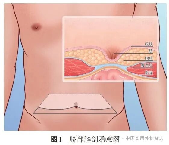
脐位于腹壁正中线,是胚胎期腹壁最晚闭合的区域。由于脐部缺乏脂肪组织,致使皮肤、筋膜及腹膜直接连接,成为腹壁最薄弱的部位之一(图1)。白线的腱膜环绕脐形成脐环。在人体胎儿时期,通过脐环部位有2条动脉、1条静脉、卵黄管及脐尿管与母体相连,以获取营养。在胎儿出生前后,上述结构逐渐闭锁,脐环闭锁后形成脐凹陷。腹腔内容物易在此薄弱部位突出形成脐疝。  随着研究深入,脐疝的定义也在不断发生改变。2020年《欧洲疝学会和美国疝学会脐疝与上腹壁疝的治疗指南》(以下简称欧美指南)认为,缺损位于脐环内腹中线的原发性疝为脐疝[2]。部分脐疝可无任何症状,病人主诉运动或体位变化等导致腹内压增加时,可出现脐部或上腹壁无痛性包块;少部分脐疝病人表现出疼痛及疼痛导致的活动受限,极少部分病人可出现嵌顿,表现为无法复位的隆起伴活动性呕吐、心动过速和低血压;严重者可进展为绞窄性疝[3-4]。
2 |病因
婴儿脐疝是由于脐环闭锁不全或延迟闭锁,胎儿出生后出现畸形,形成脐疝。成人脐疝的发病与脐环组织薄弱及腹腔压力增高有关,常见危险因素包括肥胖、代谢综合征、腹腔积液、妊娠、腹直肌分离、结肠憩室、某些结缔组织疾病和种族背景等[5-8]。
3 |诊断和鉴别诊断
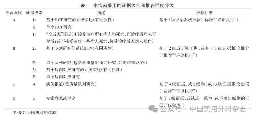
大多数脐疝病人可依据病史和体格检查确诊。典型表现为脐部可复性、质软的包块,常在病人站立、咳嗽或腹内压增加时出现。体格检查可触及脐部薄弱及缺损的边缘。对于诊断不明确者,可辅助行腹部超声、CT或MRI检查以明确诊断。腹腔镜手术时偶可发现无症状的脐疝。术前行超声、CT或MRI检查可判断脐疝缺损大小及周围器官情况[9-10]。 脐疝的诊断主要依据病史和体格检查。在诊断不明确的情况下及术前准备时,建议行超声或CT检查(证据级别:4级;推荐强度:C)。 脐疝需与上腹壁疝、脐旁疝及腹白线疝相鉴别。其中,上腹壁疝指缺损中心位于脐环与剑突之间的腹中线处的原发性疝[2];脐旁疝的定义尚不明确,有些脐周围发生的疝被报道为脐旁疝;发生于腹部正中线(腹白线)处的疝为腹白线疝,腹白线疝包含脐疝、上腹壁疝、部分缺损在白线处的脐旁疝。
4 |分型
脐疝的分型尚无统一的标准,切口疝及原发性腹壁疝的分级标准并不适合脐疝[11-12]。参考欧美指南,根据脐疝的直径将其分为小型疝(缺损直径为0~1 cm)、中型疝(>1 cm~4 cm)和大型疝(>4 cm)3个类型(证据级别:4级,推荐强度:C)。 脐疝缺损测量方法尚无统一标准,测量方法主要包括术前CT、术中腹部无充气状态测量、术中腹腔充气至15 mmHg(1 mmHg=0.133 kPa)经腹腔外测量、术中腹腔充气至15 mmHg经腹腔内测量和临床体检等。但这些测量方法得到的脐疝缺损值差异较大。术前CT测量的干扰因素相对少,且简单、可行性高,可用于术前评估(证据级别:4级,推荐强度:C)。目前,仍需进一步研究探索何种测量方法更利于指导补片的选择[10]。
5 |手术治疗
目前,手术治疗成人脐疝唯一的治愈方法。脐疝病人如出现影响生活质量、脐疝伴随症状明显、疝环直径≥2 cm、有嵌顿史、CT检查显示有肠管进入疝囊,以及病人对美观要求等,建议积极手术治疗(证据级别:4级,推荐强度:C)。手术方式包括开放手术和腹腔镜手术。根据是否使用补片,可分为单纯缝合修补和补片加强修补两种方式。 腹腔镜脐疝修补术的优势包括:(1)手术创伤小,术后疼痛轻、恢复快。(2)手术部位事件(surgical site occurrence,SSO)发生率低,术区远离脐部,术后切口感染、血肿及血清肿发生率低。(3)可同时处理腹腔其他病变。(4)在肠管或网膜组织嵌顿的情况下,有助于判断嵌顿肠管或网膜组织是否坏死。(5)保留脐部结构,满足美观要求。(6)腹腔镜手术对于保持脐环这一重要结构较开放手术更具优势。腹腔镜手术尤其适用于肥胖、糖尿病、复发疝、大疝囊病人[13-20]。欧美指南提出,对于缺损直径>4 cm或切口感染风险高的脐疝和上腹壁疝病人,推荐选择腹腔镜疝修补术[2]。结合我国临床实践,本指南建议,对于缺损直径>1 cm的中或大型脐疝可考虑腹腔镜手术修补(证据级别:2b级,推荐强度:B);对于缺损直径≤1 cm的小型脐疝,可与病人沟通后决定手术方式。 5.1 单纯缝合修补 缝合材料和缝合方式选择可能是脐疝术后复发的重要因素。常用的缝合材料为不可吸收缝合线、慢吸收缝合线和快吸收缝合线等。快吸收缝线不推荐用于任何类型的腹壁疝修补手术。缝合方式包括间断缝合、连续缝合及一些特殊的缝合方式,如Mayo技术、Keel技术等[21-23]。部分研究结果表明,无论使用哪种缝合方式,单纯缝合修补术后脐疝复发率差异均无统计学意义[24]。本指南推荐,脐疝单纯缝合修补时使用慢吸收缝线或不可吸收缝线,选择间断缝合或连续缝合方式均可(证据级别:4级,推荐强度:C)。 有研究结果显示,对缺损直径<2 cm的腹壁疝行单纯缝合修补并不增加术后疝复发率,而且可以减少手术相关并发症[25-30]。但也存在不同的研究结果,一项区域队列研究结果提示,对于小型腹壁疝,单纯缝合修补术后疝的复发率明显高于补片修补[24,31]]。此外,单纯缝合修补不适合用于同时存在腹直肌分离或既往有其他部位疝的病人[26-27]。 关于缺损直径≤1 cm腹壁疝的相关研究较少。有文献报道,即使是小型(缺损直径≤1 cm)脐疝,补片修补术也有助于降低术后复发率[32]。同样,另一研究结果也指出,对缺损直径<1 cm的脐疝采用补片修补和单纯缝合修补均安全有效;补片修补可能降低复发率,且并不增加术后感染和疼痛的风险[33]。 欧美指南推荐,小脐疝可以根据病人自身情况及意愿综合协商后决定[1]。本指南建议,对于无症状的小型脐疝可采用单纯缝合修补或补片修补,根据病人自身情况及意愿综合协商后决定;手术时保留脐环的解剖结构至关重要(证据级别:4级,推荐强度:C)。 5.2 脐疝补片修补 各研究中由于随访方法不同,脐疝术后复发率差异较大,为1%~54.5%[34-37]。多项研究结果表明,补片加强修补能显著降低脐疝术后复发率,且不增加切口感染、血清肿、血肿及慢性疼痛等并发症[38-43]。因此,对于中、大型脐疝修补手术推荐常规使用补片(证据级别:2a,推荐强度:B)。 5.3 脐疝缺损关闭 脐疝缺损通常在术中被关闭[22,44]。但在肌间补片修补及某些预制补片修补时,由于补片固定在缺损的边缘,故不关闭缺损[36,45]。腹膜前补片修补时,关闭疝缺损可防止补片移位至皮下[2]。使用补片行开放腹膜前脐疝修补时,建议使用不可吸收线或慢吸收线关闭缺损(证据级别:2a级,推荐强度:B)。 腹腔镜脐疝修补手术常采用腹腔内补片修补术(intraperitoneal onlay mesh,IPOM)。经典IPOM不关闭腹壁缺损[46]。术后可出现突起或膨出,类似于疝复发。有研究结果显示,此类突起或膨出的发生率高达31.5%[47-50]。而且,补片以桥接方式覆盖缺损,术后血清肿、感染和病人不适感的发生率较高[49-51]。若在腹腔镜IPOM术中同时关闭疝缺损,该技术称为“IPOM plus”,可重建正常、有功能的腹壁并预防腹壁突起或膨出、减少血清肿的发生[47,49-54]。对于腹腔镜肌后修补(Sublay)等术式,也建议术中关闭疝缺损,其中代表术式如Belyansky等[55]提出的增强视野全腹膜外修补术(enhanced-view totally extraperitoneal,e-TEP),以及Li等[56]提出的全腹腔镜肌后腹膜外补片修补术(totally endoscopic sublay,TES)。疝缺损关闭的方法主要为腹壁全层间断缝合和倒刺线连续缝合。此两种方法均采用不可吸收线或慢吸收线。目前尚未见两种方法的对比研究。本指南推荐采用不可吸收线或慢吸收线关闭疝缺损(证据级别:3a级,推荐强度:B)。 5.4 补片的置入层次 脐疝补片可放置于肌筋膜前(Onlay)、腹直肌间(Inlay)、腹直肌后方或腹膜外间隙(Sublay)和腹腔内(intraperitoneal)[57-59]。在没有腹直肌后鞘的区域,Sublay所指的肌后间隙等同于腹膜外间隙,而在有后鞘的中线区域,Sublay又可分为肌后-鞘前间隙(retrorectus space)和鞘后-腹膜前间隙(preperitoneal space)。 Onlay手术在缺损直径≤2 cm的脐疝病人中具有一定优势,特别是肝硬化或腹膜透析存在腹腔积液的病人。疝囊回纳后直接关闭缺损,再予补片加强修补,可以尽可能地保持腹膜完整性,避免腹腔积液渗漏,达到较好的治疗效果[59]。 文献报道,Inlay手术可能导致较高的复发率,故不建议采用[29];其他补片放置方式的复发率差异无统计学意义[60-61]。开放手术中将补片置于腹腔内不仅难以展平补片,还可能导致肠梗阻等严重并发症[62]。本指南推荐开放手术中将补片放置在腹膜前间隙(证据级别:3b级,推荐强度:B)。 腹腔镜脐疝修补术常采用IPOM,将补片置于腹腔内(证据级别:1c级,推荐强度:A)。近年来,如Sublay等新型修补方式应用逐渐增多,其将补片置于腹直肌后-鞘前间隙或鞘后-腹膜前间隙[55-56,63-65]。然而,将补片置于肌后间隙,需要采用跨中线分离(Crossover)技术将两侧后鞘内侧缘离断从而连通两侧肌后间隙,对于原发性脐疝,此方法的创伤大,破坏了腹壁原有结构[55-56,63]。与之相比,无需破坏后鞘的腹膜外放置补片的方式,理论上更适合脐疝这类原发性腹壁疝,包括尾侧入路和头侧入路的内镜下全腹膜外疝修补术(endoscopic totally extraperitoneal approach,TEA)、腹腔镜或机器人经腹腹膜前疝修补术(transabdominal preperitoneal repair, TAPP)等[64-65]。上述修补方式可使用聚丙烯补片,且避免了补片对腹腔组织器官的接触性损伤,降低术后并发症发生率及脐疝复发率,但需要更多高质量的临床试验论证,且术者学习曲线较长,建议在技术成熟的医疗单位开展[66-71](证据级别:4级,推荐强度:C)。 5.5 补片覆盖的范围 补片覆盖仅超过缺损边缘1 cm可能导致术后复发率升高,而超过缺损边缘3~5 cm则可降低复发率[22,72]。开放脐疝修补手术行腹膜前补片修补时,推荐补片覆盖超过缺损边缘3 cm(证据级别:3a级,推荐强度:B)。但对于缺损直径<1 cm的小型脐疝和上腹壁疝,补片是否也需要覆盖超过缺损边缘3 cm,目前尚缺乏明确证据支持。 腹腔镜IPOM将补片置于腹腔内,腹腔镜Sublay手术将补片置于腹直肌后鞘前,推荐补片覆盖超过缺损边缘5 cm(证据级别:3a级,推荐强度:B)。但在腹腔镜Sublay手术中,由于上腹壁贴近肋缘附近、双侧腹直肌后鞘外侧的腹膜前间隙狭窄,分离过程容易导致腹膜破损。故推荐,在腹腔镜腹膜前脐疝修补术中,补片覆盖超过缺损边缘3~5 cm(证据级别:3a级,推荐强度:B)。 5.6 补片材料选择及固定方法 开放腹膜前脐疝修补术可选用平片、预制补片或网塞等材料(证据级别:4级,推荐强度:C),目前尚无定论是否需要固定补片[2,22,68]。临床实践中,由于腹膜前间隙较疏松,可以用纱布等钝性分离拓展足够的空间,故脐疝手术切口通常较小。此时固定补片的边缘存在一定困难。推荐腹膜前脐疝修补术中无需固定补片边缘(证据级别:4级,推荐强度:C)。 腹腔镜IPOM需将补片置于腹腔内,建议使用防粘连补片(证据级别: 1c级,推荐强度:A),并采用钛钉、可吸收钉、缝合、钛钉或可吸收钉联合缝合的方式固定补片边缘[73-76] (证据级别: 4级,推荐强度:C)。选择可吸收钉可能有助于减轻术后早期疼痛。 国外文献报道,腹腔镜脐疝修补术中使用纤维蛋白胶固定补片,但国内尚未见此类产品[77-78]。随访结果显示,单纯使用纤维蛋白胶固定补片的病人术后1年的疝复发率较高[78]。
6 |特殊类型的脐疝修补策略
6.1 无症状的脐疝治疗策略 对无症状的脐疝病人可采取“观察等待”策略,不进行外科手术,在短期内被认为相对安全[79-80]。但最终19%~24%的病人接受手术治疗,其中4%的病人进行急诊手术。行急诊手术脐疝病人的病死率、再次手术率及再次入院率明显升高[80-81]。本指南专家组认为,对于无症状的脐疝,如病人无明显手术禁忌证,又有明确手术意愿,建议行外科手术治疗(证据级别:2b级,推荐强度:B)。 6.2 嵌顿及绞窄性脐疝的治疗策略 嵌顿及绞窄性脐疝需行急诊手术。清洁切口、清洁-污染切口情况下,补片修补术已被证实是安全的,故推荐使用补片[82-85](证据级别:3b级,推荐强度:B)。对于绞窄疝,不建议使用合成补片。欧美指南推荐,污染切口情况下,脐疝缺损直径<3 cm时,建议行一期缝合修补;当直接缝合修补不满意时,可考虑加用生物补片[86]。
7 |围手术期管理
7.1 择期脐疝手术的术前准备 老年脐疝病人术前行肺功能检测和心脏超声检查,侧重于改善有肺部疾病病人的氧合情况;术前戒烟时间>3周;肥胖病人须减重[26,87];糖尿病病人须控制血糖;营养状态差者须改善营养状况;存在感染者须控制感染[88-92]。对于肝硬化合并脐疝的病人,围手术期控制腹腔积液是决定手术修补时机和术后预后的关键因素[93-95]。积极治疗肝脏原发病,改善肝功能,择期手术病人的肝功能Child-Pugh分级应控制在A/B级。加强利尿,改善营养状况,减少腹腔积液产生。如存在顽固性大量腹腔积液,建议术前和术后持续行腹腔引流,以保证补片与组织间的融合。对于疝缺损较大病人,术后腹带加压包扎约2周。推荐行择期手术的脐疝病人术前积极戒烟、减重、控制腹腔积液(证据级别:4级,推荐强度:C),术前禁食12 h及进行肠道准备。 7.2 择期脐疝手术的麻醉选择及抗生素使用 依据美国麻醉医师协会(American Society of Anesthesiologists,ASA)手术风险评估标准,评估机体是否可以耐受手术。采用气管插管或静脉复合全身麻醉,依据《抗菌药物药代动力学/药效学临床应用专家共识》,Ⅰ类清洁切口,仅在预期会进行广泛的粘连松解,并根据手术复杂性和持续时间,置入补片时可预防使用抗菌药物[96]。对于需植入补片的脐疝手术,建议术前常规预防性使用抗菌药物(证据级别:4级,推荐强度:C)。手术时间>3 h,术野污染严重,或存在特殊情况(如糖尿病、肝硬化、使用免疫抑制剂等)病人,围手术期建议预防性和治疗性使用抗生素。
8 |日间手术的麻醉方式选择
脐疝病人在局部麻醉下行修补手术安全可行,且可以缩短住院时间[97-99]。研究结果显示,采用局部麻醉并未较全身麻醉方式显著增加围手术期并发症、过敏反应及麻醉相关死亡风险[2,100]。腹横肌、腹直肌鞘神经阻滞可提供更好的镇痛效果[101-102]。但并无研究结果支持局部麻醉优于全身麻醉,也没有相关共识推荐使用何种局麻药物和技术。而且,全身麻醉可使腹壁处于松弛状态,有利于手术修补,减少术后复发。 本指南推荐,日间脐疝手术病人可选择局部麻醉(证据级别:3b级,推荐强度:B);条件允许时可联合应用下腹壁神经阻滞,以增强术中镇痛效果(证据级别:3b级,推荐强度:B)。
9 |机器人辅助脐疝修补术
目前,机器人辅助脐疝修补术开展逐渐增多。机器人手术可更便捷地完成缺损关闭,并将补片置于腹直肌后间隙[103-108]。其优点包括创伤小、住院时间短、术中缝合便利、减少术后疼痛等,但亦存在手术时间和学习曲线长等缺点。术后并发症与开放手术和腹腔镜手术相近。该技术应在具备相关资质的医疗机构中谨慎开展(证据级别:4级,推荐强度:C)。 本指南不仅包含了脐疝的定义、病因、诊断和鉴别诊断、分类、手术治疗及围手术期管理的各个细节,也涵盖了无症状脐疝、嵌顿及绞窄性脐疝等特殊类型的脐疝修补策略,以及机器人辅助脐疝修补术等新兴的治疗方法。期望本指南为广大疝与腹壁外科医师更规范地开展脐疝手术提供依据,未来也将根据更多的临床证据及专家经验进行更新。  参考文献 (在框内滑动手指即可浏览) [1] Burcharth J, Pedersen MS, Pommergaard HC, et al. The prevalence of umbilical and epigastric hernia repair: A nationwide epidemiologic study[J]. Hernia, 2015, 19(5):815-819. DOI:10.1007/s10029-015-1376-3. [2] European Hernia Society, Americas Hernia Society. Guidelines for treatment of umbilical and epigastric hernias from the European Hernia Society and Americas Hernia Society[J]. Br J Surg, 2020, 107(3):171-190. DOI:10.1002/bjs.11428. [3] Hoffmann H, Mechera R, Nowakowski D, et al. Gender differences in epigastric hernia repair: A propensity score matching analysis of 15,925 patients from the Herniamed registry[J]. Hernia, 2023, 27(4):829-838. DOI:10.1007/s10029-023-02799-8. [4] Patel S, Smiley A, Feingold C, et al. Chances of mortality are 3.5-times greater in elderly patients with umbilical hernia than in adult patients: an analysis of 21,242 patients[J]. Int J Environ Res Public Health, 2022, 19(16):10402. DOI:10.3390/ijerph191610402. [5] Nishihara Y, Asami M, Shimada T, et al. Comorbid rectus abdominis diastasis is a risk factor for recurrence of umbilical hernia in Japanese patients[J]. Asian J Endosc Surg, 2021, 14(3):368-372. DOI:10.1111/ases.12868. [6] Henriksen NA. Systemic and local collagen turnover in hernia patients[J]. Dan Med J, 2016, 63:B5265. PMID: 27399987. [7] Oma E, Jorgensen LN, Meisner S, et al. Colonic diverticulosis is associated with abdominal wall hernia[J]. Hernia, 2017, 21(4):525-529. DOI: 10.1007/s10029-017-1598-7. [8] Kelly KB, Ponsky TA. Pediatric abdominal wall defects[J]. Surg Clin North Am, 2013, 93(5):1255-1267. DOI: 10.1016/j.suc.2013.06.016. [9] 陈健, 王山, 云香. 多层螺旋CT联合MRI在腹壁疝患者中的诊断效果及临床治疗研究[J]. 中华疝和腹壁外科杂志(电子版), 2021, 15 (3): 255-258. DOI: 10.3877/cma.j.issn.1674-392X.2021.03.011. [10] Cherla DV, Lew DF, Escamilla RJ, et al. Differences of alternative methods of measuring abdominal wall hernia defect size: A prospective observational study[J]. Surg Endosc, 2018, 32(3):1228-1233. DOI: 10.1007/s00464-017-5797-1. [11] 中华医学会外科学分会疝与腹壁外科学组, 中国医师协会外科医师分会疝和腹壁外科专家工作组. 腹壁切口疝诊疗指南(2024版)[J]. 中国实用外科杂志, 2025, 45(2):128-136. DOI:10.19538/j.cjps.issn1005-2208.2025.02.02. [12] 中华医学会外科学分会疝与腹壁外科学组, 中华医学会外科学分会腹腔镜与内镜外科学组. 成人原发性腹壁疝腹腔镜手术中国专家共识(2025版)[J]. 中国实用外科杂志, 2025, 45(1):25-33. [13] Hajibandeh S, Hajibandeh S, Sreh A, et al. Laparoscopic versus open umbilical or paraumbilical hernia repair: A systematic review and Meta-analysis[J]. Hernia, 2017, 21(6):905-916. DOI: 10.1007/s10029-017-1683-y. [14] Castro PM, Rabelato JT, Monteiro GG, et al. Laparoscopy versus laparotomy in the repair of ventral hernias: Systematic review and Meta-analysis[J]. Arq Gastroenterol, 2014, 51(3):205-211. DOI: 10.1590/s0004-2803201400030008. [15] Zhang Y, Zhou H, Chai Y, et al. Laparoscopic versus open incisional and ventral hernia repair: A systematic review and Meta-analysis[J]. World J Surg, 2014, 38(9):2233-2240. DOI: 10.1007/s00268-014-2578-z. [16] Henriksen NA, Jorgensen LN, Friis-Andersen H, et al. Open versus laparoscopic umbilical and epigastric hernia repair: nationwide data on short- and long-term outcomes[J]. Surg Endosc, 2022, 36(1):526-532. DOI:10.1007/s00464-021-08312-5. [17] Williams KN, Hussain L, Fellner AN, et al. Updated outcomes of laparoscopic versus open umbilical hernia repair in patients with obesity based on a National Surgical Quality Improvement Program review[J]. Surg Endosc, 2020, 34(8):3584-3589. DOI:10.1007/s00464-019-07129-7. [18] Fafaj A, Tastaldi L, Alkhatib H, et al. Is there an advantage to laparoscopy over open repair of primary umbilical hernias in obese patients? An analysis of the Americas Hernia Society Quality Collaborative (AHSQC)[J]. Hernia, 2021, 25(3):579-585. DOI:10.1007/s10029-020-02218-2. [19] Elhage SA, Pflederer CT, Ayuso SA, et al. Multicenter analysis of laparoscopic versus open umbilical hernia repair with mesh: outcomes and quality of life (QoL)[J]. Surg Endosc, 2022, 36(9):6822-6831. DOI:10.1007/s00464-021-08971-4. [20] Shao JM, Elhage SA, Prasad T, et al. Outcomes of laparoscopic-assisted, open umbilical hernia repair[J]. Am Surg, 2020, 86(8):1001-1004. DOI:10.1177/0003134820942162. [21] Tunio NA. Hernioplasty: tension free mesh repair versus Mayos repair for umbilical hernias[J]. J Pak Med Assoc, 2017, 67(1):24-26. [22] Kaufmann R, Halm JA, Eker HH, et al. Mesh versus suture repair of umbilical hernia in adults: A randomised, double-blind, controlled, multicentre trial[J]. Lancet, 2018, 391(10123):860-869. DOI: 10.1016/S0140-6736(18)30298-8. [23] Mayo WJ. An operation for the radical cure of umbilical hernia[J]. Ann Surg, 1901, 34(2):276-280. DOI: 10.1097/00000658-190107000-00021. [24] Christoffersen MW, Helgstrand F, Rosenberg J, et al. Long-term recurrence and chronic pain after repair for small umbilical or epigastric hernias: A regional cohort study[J]. Am J Surg, 2015, 209(4):725-732. DOI: 10.1016/j.amjsurg.2014.05.021. [25] Dalenbäck J, Andersson C, Ribokas D, et al. Long-term follow-up after elective adult umbilical hernia repair: low recurrence rates also after non-mesh repairs[J]. Hernia, 2013, 17(4):493-497. DOI: 10.1007/s10029-012-0988-0. [26] Köhler G, Luketina RR, Emmanuel K. Sutured repair of primary small umbilical and epigastric hernias: Concomitant rectus diastasis is a significant risk factor for recurrence[J]. World J Surg, 2015, 39(1):121-126. DOI: 10.1007/s00268-014-2765-y. [27] Winsnes A, Haapamäki MM, Gunnarsson U, et al. Surgical outcome of mesh and suture repair in primary umbilical hernia: postoperative complications and recurrence[J]. Hernia, 2016, 20(4):509-516. DOI: 10.1007/s10029-016-1466-x. [28] Christoffersen MW, Helgstrand F, Rosenberg J, et al. Lower reoperation rate for recurrence after mesh versus sutured elective repair in small umbilical and epigastric hernias. A nationwide register study[J]. World J Surg, 2013, 37(11):2548-2552. DOI: 10.1007/s00268-013-2160-0. [29] Kang E, Collins JC. Primary umbilical hernia repair: A large-volume single-surgeon study[J]. Am Surg, 2019, 85(10):1159-1161. [30] Mitura K, Skolimowska-Rzewuska M, Rzewuska A, et al. Is mesh always necessary in every small umbilical hernia repair? Comparison of standardized primary sutured versus patch repair: Retrospective cohort study[J]. Hernia, 2021, 25(3):571-577. DOI:10.1007/s10029-020-02170-1. [31] Köckerling F, Brunner W, Mayer F, et al. Assessment of potential influencing factors on the outcome in small (<2 cm) umbilical hernia repair: A registry-based multivariable analysis of 31,965 patients[J]. Hernia, 2021, 25(3):587-603. DOI:10.1007/s10029-020-02305-4. [32] Henriksen NA, Jensen KK, Bisgaard T, et al. Suture or mesh repair of the smallest umbilical hernias: A nationwide database study[J]. World J Surg, 2022, 46(8):1898-1905. DOI:10.1007/s00268-022-06520-1. [33] Frey S, Beauvais A, Soler M, et al. Suture versus open mesh repair for small umbilical hernia: results of a propensity-matched cohort study[J]. Surgery, 2023, 174(3):593-601. DOI:10.1016/j.surg.2023.05.015. [34] Halm JA, Heisterkamp J, Veen HF, et al. Long-term follow-up after umbilical hernia repair: are there risk factors for recurrence after simple and mesh repair[J]. Hernia, 2005, 9(3):334-337. DOI: 10.1007/s10029-005-0010-1. [35] Arroyo A, García P, Pérez F, et al. Randomized clinical trial comparing suture and mesh repair of umbilical hernia in adults[J]. Br J Surg, 2001, 88(10):1321-1323. DOI: 10.1046/j.0007-1323.2001.01893.x. [36] Polat C, Dervisoglu A, Senyurek G, et al. Umbilical hernia repair with the prolene hernia system[J]. Am J Surg, 2005, 190(1):61-64. DOI: 10.1016/j.amjsurg.2004.09.021. [37] Katzen MM, Sacco J, Ku D, et al. Hernia recurrence after primary repair of small umbilical hernia defects[J]. Am J Surg, 2022, 224(6):1357-1361. DOI:10.1016/j.amjsurg.2022.09.019. [38] Tao Z, Ordonez J, Huerta S. Hernia size and mesh placement in primary umbilical hernia repair[J]. Am Surg, 2021, 87(6):1005-1013. DOI:10.1177/0003134820971624. [39] Kaufmann R, Halm JA, Lange JF. The power of level-1 evidence in umbilical hernia repair: Mesh repair is supported by strong evidence[J]. Hernia, 2021, 25(1):247-248. DOI:10.1007/s10029-020-02185-8. [40] Shrestha D, Shrestha A, Shrestha B. Open mesh versus suture repair of umbilical hernia: Meta-analysis of randomized controlled trials[J]. Int J Surg, 2019, 62:62-66. DOI: 10.1016/j.ijsu.2018.12.015. [41] Porrero JL, Cano-Valderrama O, Villar S, et al. Umbilical hernia repair with composite prosthesis: A single-centre experience[J]. Hernia, 2019, 23(1):143-147. DOI: 10.1007/s10029-018-1843-8. [42] Aslani N, Brown CJ. Does mesh offer an advantage over tissue in the open repair of umbilical hernias? A systematic review and meta-analysis[J]. Hernia, 2010, 14(5):455-462. DOI: 10.1007/s10029-010-0705-9. [43] Malibary N, Shurrab M, Albariqi MO, et al. Quality of life after umbilical hernia repair[J]. Cureus, 2021, 13(10):e19016. DOI:10.7759/cureus.19016. [44] Haskins IN, Voeller GR, Stoikes NF, et al. Onlay with adhesive use compared with sublay mesh placement in ventral hernia repair: was Chevrel right? An Americas Hernia Society quality collaborative analysis[J]. J Am Coll Surg, 2017, 224(5):962-970. DOI: 10.1016/j.jamcollsurg.2017.01.048. [45] Martin DF, Williams RF, Mulrooney T, et al. Ventralex mesh in umbilical/epigastric hernia repairs: clinical outcomes and complications[J]. Hernia, 2008, 12(4):379-383. DOI: 10.1007/s10029-008-0351-7. [46] LeBlanc KA, Booth WV. Avoiding complications with laparoscopic herniorrhaphy[J]. Surg Laparosc Endosc, 1993, 3(5):420-424. [47] Nguyen DH, Nguyen MT, Askenasy EP, et al. Primary fascial closure with laparoscopic ventral hernia repair: systematic review[J]. World J Surg, 2014, 38(12):3097-3104. DOI: 10.1007/s00268-014-2722-9. [48] Lambrecht JR, Vaktskjold A, Trondsen E, et al. Laparoscopic ventral hernia repair: outcomes in primary versus incisional hernias: no effect of defect closure[J]. Hernia, 2015, 19(3):479-486. DOI: 10.1007/s10029-015-1345-x. [49] Suwa K, Okamoto T, Yanaga K. Closure versus non-closure of fascial defects in laparoscopic ventral and incisional hernia repairs: A review of the literature[J]. Surg Today, 2016, 46(7):764-773. DOI: 10.1007/s00595-015-1219-y. [50] Tandon A, Pathak S, Lyons NJ, et al. Meta-analysis of closure of the fascial defect during laparoscopic incisional and ventral hernia repair[J]. Br J Surg, 2016, 103(12):1598-1607. DOI: 10.1002/bjs.10268. [51] Hauters P, Desmet J, Gherardi D, et al. Assessment of predictive factors for recurrence in laparoscopic ventral hernia repair using a bridging technique[J]. Surg Endosc, 2017, 31(9):3656-3663. DOI: 10.1007/s00464-016-5401-0. [52] Martin-del-Campo LA, Miller HJ, Elliott HL, et al. Laparoscopic ventral hernia repair with and without defect closure: comparative analysis of a single-institution experience with 783 patients[J]. Hernia, 2018, 22(6):1061-1065. DOI: 10.1007/s10029-018-1812-2. [53] Christoffersen MW, Westen M, Rosenberg J, et al. Closure of the fascial defect during laparoscopic umbilical hernia repair: a randomized clinical trial[J]. Br J Surg, 2020, 107(3):200-208. DOI:10.1002/bjs.11490. [54] Khan S, Siddiqa M, Rehman HU. Comparison of frequency of seroma formation in laparoscopic para umbilical hernia repair with and without primary closure of defect[J]. J Pak Med Assoc, 2022, 72(2):265-269. DOI:10.47391/JPMA.1487. [55] Belyansky I, Daes J, Radu VG, et al. A novel approach using the enhanced-view totally extraperitoneal (eTEP) technique for laparoscopic retromuscular hernia repair[J]. Surg Endosc, 2018, 32(3):1525-1532. DOI: 10.1007/s00464-017-5840-2. [56] Li B, Qin C, Bittner R. Totally endoscopic sublay (TES) repair for midline ventral hernia: surgical technique and preliminary results[J]. Surg Endosc, 2018, 32(12):5054-5060. DOI:10.1007/s00464-018-6568-3. [57] Muysoms F, Jacob B. International Hernia Collaboration consensus on nomenclature of abdominal wall hernia repair[J]. World J Surg, 2018, 42(2):302-304. DOI: 10.1007/s00268-017-4115-3. [58] Muysoms F. IPOM: history of an acronym[J]. Hernia, 2018, 22(5):743-746. DOI: 10.1007/s10029-018-1804-2. [59] Melkemichel M, Stjärne L, Bringman S, et al. Onlay mesh repair for treatment of small umbilical hernias ≤2 cm in adults: a single-centre investigation[J]. Hernia, 2022, 26(6):1483-1489. DOI:10.1007/s10029-021-02509-2. [60] 刘小莉, 马秋月, 朱熠林. 成人脐疝修补术式的临床应用探讨[J]. 中华疝和腹壁外科杂志(电子版), 2023, 16(3):269-273. DOI:10.3877/cma.j.issn.1674-392X.2023.03.007. [61] Person H, Mojallal A, Braye F, et al. Techniques of concomitant abdominoplasty and umbilical hernia repair: A review[J]. Aesthet Surg J, 2021, 41(7):NP831-NP839. DOI:10.1093/asj/sjab094. [62] Keating JJ, Kennedy GT, Datta J, et al. Outcomes of 157 V-PatchTM implants in the repair of umbilical, epigastric, and incisional hernias[J]. Am Surg, 2016, 82(1):6-10. PMID:26802839. [63] Schwarz J, Reinpold W, Bittner R. Endoscopic mini/less open sublay technique (EMILOS) - a new technique for ventral hernia repair[J]. Langenbecks Arch Surg, 2017, 402(1):173-180. DOI: 10.1007/s00423-016-1522-0. [64] Li B, Qin C, Bittner R. Endoscopic totally extraperitoneal approach (TEA) technique for primary ventral hernia repair[J]. Surg Endosc,2020,34(8):3734-3741. DOI:10.1007/s00464-020-07575-8. [65] Li B, Qin C, Liu D, et al. Subxiphoid top-down endoscopic totally preperitoneal approach (eTPA) for midline ventral hernia repair[J]. Langenbecks Arch Surg,2021,406(6):2125-2132. DOI:d10.1007/s00423-021-02259-w. [66] 赵学飞, 靳翠红, 申英末. 全腹腔镜Sublay 手术与腹腔镜IPOM手术治疗脐疝的回顾性分析[J]. 中华疝和腹壁外科杂志(电子版), 2025, 19(1): 51-55. DOI: 10.3877/cma.j.issn.1674-392X.2025.01.010. [67] Belyansky I, Reza Zahiri H, Sanford Z, et al. Early operative outcomes of endoscopic (eTEP access) robotic-assisted retromuscular abdominal wall hernia repair[J]. Hernia, 2018, 22(5):837-847. DOI: 10.1007/s10029-018-1795-z. [68] Nevo N, Goldstein AL, Staierman M, et al. eTEP inferior access with tailored retromuscular dissection for small to mid-sized umbilical hernia repair with or without inguinal hernia: early experience[J]. Hernia, 2022, 26(6):1491-1499. DOI:10.1007/s10029-022-02649-z. [69] Zhang Z, Li L, Liu B, et al. Effect of laparoscopic total extraperitoneal umbilical hernia repair on incision infection, complication rate, and recurrence rate in patients with umbilical hernia[J]. J Healthc Eng, 2022, 2022:7055045. DOI:10.1155/2022/7055045. [70] Cai XY, Chen K, Pan Y, et al. Total endoscopic sublay mesh repair for umbilical hernias[J]. Medicine (Baltimore), 2021, 100(25): e26334. DOI:10.1097/MD.0000000000026334. [71] Kumar N, Jaiswal P, Sinha N, et al. Novel technique of laparoscopic e-TEP (extended view totally extraperitoneal repair) for umbilical hernia at a tertiary care centre of Eastern India: A case series[J]. Maedica (Bucur), 2022, 17(2):329-335. DOI:10.26574/maedica.2022.17.2.329. [72] LeBlanc KA. Proper mesh overlap is a key determinant in hernia recurrence following laparoscopic ventral and incisional hernia repair[J]. Hernia, 2016, 20(1):85-99. DOI: 10.1007/s10029-015-1399-9. [73] Guérin G, Turquier F. Impact of the defect size, the mesh overlap and the fixation depth on ventral hernia repairs: A combined experimental and numerical approach[J]. Hernia, 2013, 17(5):647-655. DOI: 10.1007/s10029-013-1050-6. [74] Reynvoet E, Deschepper E, Rogiers X, et al. Laparoscopic ventral hernia repair: is there an optimal mesh fixation technique? A systematic review[J]. Langenbecks Arch Surg, 2014, 399(1):55-63. DOI: 10.1007/s00423-013-1126-x. [75] Khan RMA, Bughio M, Ali B, et al. Absorbable versus non-absorbable tacks for mesh fixation in laparoscopic ventral hernia repair: A systematic review and Meta-analysis[J]. Int J Surg, 2018, 53:184-192. DOI: 10.1016/j.ijsu.2018.03.042. [76] Sajid MS, Parampalli U, McFall MR. A meta-analysis comparing tacker mesh fixation with suture mesh fixation in laparoscopic incisional and ventral hernia repair[J]. Hernia, 2013, 17(2):159-166. DOI: 10.1007/s10029-012-1017-z. [77] Eriksen JR, Bisgaard T, Assaadzadeh S, et al. Randomized clinical trial of fibrin sealant versus titanium tacks for mesh fixation in laparoscopic umbilical hernia repair[J]. Br J Surg, 2011, 98(11):1537-1545. DOI: 10.1002/bjs.7646. [78] Eriksen JR, Bisgaard T, Assaadzadeh S, et al. Fibrin sealant for mesh fixation in laparoscopic umbilical hernia repair: 1-year results of a randomized controlled double-blinded study[J]. Hernia, 2013, 17(4):511-514. DOI: 10.1007/s10029-013-1101-z. [79] Kokotovic D, Sjølander H, Gögenur I, et al. Watchful waiting as a treatment strategy for patients with a ventral hernia appears to be safe[J]. Hernia, 2016, 20(2):281-287. DOI: 10.1007/s10029-016-1464-z. [80] Holihan JL, Flores-Gonzalez JR, Mo J, et al. A prospective assessment of clinical and patient-reported outcomes of initial non-operative management of ventral hernias[J]. World J Surg, 2017, 41(10):1267-1273. DOI: 10.1007/s00268-016-3859-5. [81] Strosberg DS, Pittman M, Mikami D. Umbilical hernias: the cost of waiting[J]. Surg Endosc, 2017, 31(2):901-906. DOI: 10.1007/s00464-016-5052-1. [82] Abdel-Baki NA, Bessa SS, Abdel-Razek AH. Comparison of prosthetic mesh repair and tissue repair in the emergency management of incarcerated para-umbilical hernia: A prospective randomized study[J]. Hernia, 2007, 11(2):163-167. DOI: 10.1007/s10029-007-0189-4. [83] Bessa SS, Abdel-Razek AH. Results of prosthetic mesh repair in the emergency management of the acutely incarcerated and/or strangulated ventral hernias: A seven years study[J]. Hernia, 2013, 17(1):59-65. DOI: 10.1007/s10029-012-0938-x. [84] Nieuwenhuizen J, van Ramshorst GH, ten Brinke JG, et al. The use of mesh in acute hernia: Frequency and outcome in 99 cases[J]. Hernia, 2011, 15(3):297-300. DOI: 10.1007/s10029-010-0779-4. [85] Alves JR, Spengler LFM, Justino LB, et al. Umbilical and epigastric hernia repair: A systematic review[J]. Arq Bras Cir Dig, 2024, 37:e1807. DOI:10.1590/0102-6720202400014e1807. [86] Birindelli A, Sartelli M, Di Saverio S, et al. 2017 update of the WSES guidelines for emergency repair of complicated abdominal wall hernias[J]. World J Emerg Surg, 2017, 12:37. DOI: 10.1186/s13017-017-0149-y. [87] Shankaran V, Weber DJ, Reed RL II, et al. A review of available prosthetics for ventral hernia repair[J]. Ann Surg, 2011, 253(1):16-26. DOI: 10.1097/SLA.0b013e3181f9b6e6. [88] Christoffersen MW. Clinical outcomes after elective repair for small umbilical and epigastric hernias[J]. Dan Med J, 2015, 62(4): B5161. PMID: 26522486. [89] Helgstrand F, Jørgensen LN, Rosenberg J, et al. Nationwide prospective study on readmission after umbilical or epigastric hernia repair[J]. Hernia, 2013, 17(4):487-492. DOI: 10.1007/s10029-013-1120-9. [90] DeLancey JO, Blay E Jr, Hewitt DB, et al. The effect of smoking on 30-day outcomes in elective hernia repair[J]. Am J Surg, 2018, 216(3):471-474. DOI: 10.1016/j.amjsurg.2018.03.004. [91] Donovan K, Denham M, Kuchta K, et al. Predictors for recurrence after open umbilical hernia repair in 979 patients[J]. Surgery, 2019, 166(4):615-622. DOI:10.1016/j.surg.2019.04.040. [92] Mannion J, Hamed MK, Negi R, et al. Umbilical hernia repair and recurrence: Need for a clinical trial?[J]. BMC Surg, 2021, 21(1):365. DOI:10.1186/s12893-021-01358-1. [93] Bronswijk M, Jaekers J, Vanella G, et al. Umbilical hernia repair in patients with cirrhosis: Who, when and how to treat[J]. Hernia, 2022, 26(6):1447-1457. DOI: 10.1007/s10029-022-02617-7. [94] Snitkjær C, Jensen KK, Henriksen NA, et al. Umbilical hernia repair in patients with cirrhosis: Systematic review of mortality and complications[J]. Hernia, 2022, 26(6):1435-1445. DOI:10.1007/s10029-022-02598-7. [95] Adiamah A, Rashid A, Crooks CJ, et al. The impact of urgency of umbilical hernia repair on adverse outcomes in patients with cirrhosis: A population-based cohort study from England[J]. Hernia, 2024, 28(1):109-117. DOI:10.1007/s10029-023-02898-6. [96] 中国医药教育协会感染疾病专业委员会. 抗菌药物药代动力学/药效学理论临床应用专家共识[J]. 中华结核和呼吸杂志, 2018, 41(6):409-446. DOI:10.3760/cma.j.issn.1001-0939.2018.06.004. [97] Loss L, Meier J, Phung T, et al. Feasibility of umbilical hernia repair under local anesthesia and monitored anesthesia care at a Veteran Affairs hospital[J]. Am Surg, 2022, 88(2):167-173. DOI:10.1177/0003134820951452. [98] Zuvela M, Galun D, Bogdanovic A, et al. Management of epigastric, umbilical, spigelian and small incisional hernia as a day case procedure: Results of long-term follow-up after open preperitoneal flat mesh technique[J]. Hernia, 2021, 25(5):1095-1101. DOI:10.1007/s10029-021-02446-0. [99] Jairam AP, Kaufmann R, Muysoms F, et al. The feasibility of local anesthesia for the surgical treatment of umbilical hernia: a systematic review of the literature[J]. Hernia, 2017, 21(2):223-231. DOI: 10.1007/s10029-017-1577-z. [100] Meier J, Berger M, Hogan TP, et al. Local anesthesia is associated with fewer complications in umbilical hernia repair in frail veterans[J]. J Surg Res, 2021, 266:88-95. DOI:10.1016/j.jss.2021.04.006. [101] Grape S, Kirkham KR, Albrecht E. The analgesic efficacy of transversus abdominis plane block vs. wound infiltration after inguinal and infra-umbilical hernia repairs: A systematic review and Meta-analysis with trial sequential analysis[J]. Eur J Anaesthesiol, 2022, 39(7):611-618. DOI:10.1097/EJA.0000000000001668. [102] Wang Y, Li M, Wang Z, et al. Use of bilateral rectus sheath blocks in open umbilical hernia repair[J]. Pain Physician, 2024, 27(2):E239-E244. PMID: 38324789. [103] Henriksen NA, Jensen KK, Muysoms F. Robot-assisted abdominal wall surgery: A systematic review of the literature and Meta-analysis[J]. Hernia, 2019, 23(1):17-27. DOI: 10.1007/s10029-018-1872-3. [104] Anoldo P, Manigrasso M, D'Amore A, et al. Dual docking technique for robotic repair of simultaneous inguinal and umbilical hernia: A preliminary single center experience[J]. Int J Med Robot, 2023, 19(5): e2586. DOI:10.1002/rcs.2586. [105] Sugiyama G, Chivukula S, Chung PJ, et al. Robot-assisted transabdominal preperitoneal ventral hernia repair[J]. JSLS, 2015, 19(4): e2015.00092. DOI: 10.4293/JSLS.2015.00092. [106] Kennedy M, Barrera K, Akelik A, et al. Robotic TAPP ventral hernia repair: Early lessons learned at an inner city safety net hospital[J]. JSLS, 2018, 22(4):e2017.00070. DOI: 10.4293/JSLS.2017.00070. [107] Carbonell AM, Warren JA, Prabhu AS, et al. Reducing length of stay using a robotic-assisted approach for retromuscular ventral hernia repair: A comparative analysis from the Americas Hernia Society quality collaborative[J]. Ann Surg, 2018, 267(2):210-217. DOI: 10.1097/SLA.0000000000002244. [108] Warren JA, Cobb WS, Ewing JA, et al. Standard laparoscopic versus robotic retromuscular ventral hernia repair[J]. Surg Endosc, 2017, 31(1):324-332. DOI: 10.1007/s00464-016-4975-x. (2026-03-03收稿) 版权声明 本文为《中国实用外科杂志》原创文章。其他媒体、网站、公众号等如需转载本文,请联系本刊编辑部获得授权,并在文题下醒目位置注明“原文刊发于《中国实用外科杂志》,卷(期):起止页码”。谢谢合作! 














引用 / 出处
中华医学会外科学分会疝与腹壁外科学组. 指南与共识|成人脐疝诊疗指南(2026版)[J]. 中国实用外科杂志, 2026.