优化肺功能检查多学科临床路径与管理模式专家共识
Recommendation 7:
① PFT reports from different PFT analyzers should be integrated with HIS, picture archiving and communication system (PACS) and laboratory information system (LIS) to enable automated data synchronization. (Strong Recommendation)
② Recommendations for information system access standards and conversion requirements: digital PFT reports should follow a unified and standardized format for HIS integration, including the patient's demographics, test indicators, data, images and the electronic signature of the operator or reviewing physician. If digital data cannot be integrated, images or PDFs of the reports can be uploaded to PACS. (Strong Recommendation)
Question 8: How to establish a standardized PFT data platform within healthcare institutions?
Recommendation 8:
① Integrate PFT data across all hospital departments to ensure real-time, accurate synchronization with the Hospital Information System (HIS). Records of the types, frequency and results of PFTs can be used to establish the unified, standardized PFT database for centralized management and analysis. (Strong Recommendation)
② The platform should feature basic functions such as data storage, querying, statistics and quality control. It is recommended to embed a standardized quality assessment and grading system within electronic reports to assist clinicians in rapidly evaluating data quality and interpreting the reliability of results. (Strong Recommendation)
③ Patients with confirmed chronic respiratory diseases should be integrated into the platform, or PFT data should be incorporated into existing patient management systems. This integration supports clinical research and hospital administration and enables the visual management of longitudinal pulmonary function data. (Strong Recommendation)
Question 9: How to establish a Cloud-based Data Sharing Platform?
Recommendation 9: Standards and specifications for uploading PFT data should be established to ensure data security and patient privacy. PFT data sharing among medical institutions within the region should be achieved through regional cloud storage to provide the data support for hierarchical diagnosis and treatment and telemedicine. In principle, patient information should be anonymized prior to being uploaded to regional sub-centers. Regional sub-centers are responsible for reviewing reports, revising conclusions, and assigning quality control ratings. The cloud data center should aggregate the quality control data from all sub-centers, and regularly evaluate and sample the sub-centers' quality control reports. (Strong Recommendation)
Question 10: How to improve the quality control and the interpretation of PFTs?
Recommendation 10:
① Quality control and interpretation of PFT results should be performed by physicians or technicians who have received specialized training in PFTs and possess relevant qualifications. In addition, AI can be utilized to achieve quality control and interpretation of pulmonary function test results, promoting the optimal allocation of medical resources and improving efficiency. (Strong Recommendation)
② Implementation methods for AI-assisted PFTs: PFT equipment manufacturers should integrate AI-assisted PFT systems into instruments or cloud platforms. Using AI deep learning and other models to automatically identify abnormalities, the acceptability, repeatability and quality grading of PFT results can be intelligently interpreted, more accurately distinguishing types of ventilation dysfunction, improving diagnostic accuracy and efficiency, and achieving efficient automatic identification of abnormal populations. (Strong Recommendation)
Currently, quality control and result interpretation of PFTs rely primarily on manual assessment, which is inefficient, subject to observer bias, and requires trained and qualified
· 388 ·
中华结核和呼吸杂志2026年4月第49卷第4期 Chin J Tuberc Respir Dis, April 2026, Vol. 49, No. 4
personnel. The introduction of AI technology can enable automatic quality control and assisted interpretation of PFT data, enhancing the efficiency and quality of PFTs.
AI deep learning models based on large-scale standardized PFT data can perform real-time evaluation of data collection and curve quality during the testing process, issuing timely warnings and requesting re-testing for unqualified results. Acceptable PFT results can be used for further determining the abnormalities, assessing the type and severity of pulmonary function impairment, and intelligently generating real-time warnings, patient guidance and auxiliary interpretation, improving the quality control and interpretation efficiency of PFT results.
Question 11: How to configure equipment and personnel for the PFT laboratory?
Recommendation 11:
① Space configuration: A comfortable examination environment for patients should be provided by establishing PFT suites, ensuring reasonable spatial layout with proper ventilation and temperature/humidity control. (Strong Recommendation)
② Hardware configuration: The PFT devices should be equipped according to the specific needs of the healthcare institution. The equipment should comply with industry technical specifications, undergo regular calibration and maintenance, and be documented in comprehensive equipment management logs. (Strong Recommendation)
③ Personnel configuration: The full-time technical personnel should be assigned reasonably according to the equipment and workload needs, and should undergo the relevant training and pass the assessments. (Strong Recommendation)
Question 12: How to improve the patient experience and PFT efficiency?
Recommendation 12:
① By setting up self-service terminals for PFTs or using mobile applications, the process of registration, payment, appointment, check-in, and report viewing can be completed quickly, which helps improve the patient experience, reduce waiting time, and optimize workflow. (Strong Recommendation)
② Instructional videos can visually help patients learn about the significance of PFTs, improve maneuver compliance, and adherence to precautions. (Strong Recommendation)
③ Adopting augmented reality technology (e.g., head-mounted displays) helps facilitate rapid learning and improved testing efficiency. Providing an immersive interactive experience increases patient engagement and understanding, which helps to improve the implementation rate and efficiency of PFTs. (Strong Recommendation)
Question 13: When conducting multidisciplinary PFTs, how to set the target testing rates for different departments?
Recommendation 13: In addition to routine clinical application in the Department of Pulmonary and Critical Care Medicine (PCCM), PFTs are also significant in multiple departments such as Surgery, Cardiology and Geriatrics, and should be encouraged in high-risk populations. It is recommended that the proportion of PFTs ordered by clinical departments other than PCCM Department and Thoracic Surgery Department should constitute more than 30% of the total hospital-wide PFT volume. (Strong Recommendation)
Question 14: How to set the assessment criteria for PFTs?
Recommendation 14: Competency-based criteria for physicians and technicians performing PFTs should be established, encompassing both the quantity and quality of tests. Personnel who do not meet the standards should undergo re-training and re-assessment. (Strong Recommendation)
This consensus is registered on the International Practice Guideline Registration and Transparency Platform: PREPARE-2025CN1382 DOI: 10.3760/cma.j.cn112147-20251114-00713
Fund program: National Key Research and Development Program of China (2023ZD0506300); Guangzhou National Laboratory Program (SRPG22-018,SRPG22-016)
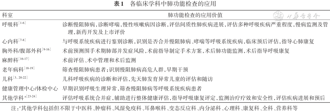
肺功能检查是呼吸系统疾病诊断、严重程度评价、治疗方案制定和效果评价以及慢病筛查、疾病随访、急性加重或发作预警等慢病管理的关键技术,也是呼吸学科的常用质控医疗指标和学科能力建设的重要一环。但除呼吸学科外,由于全身多种
疾病可伴发肺功能受损,肺功能检查在众多呼吸相关的临床学科中亦具有广泛的应用价值(表1)。
当前我国居民肺功能检查实际开展率明显偏低,2019—2020年调查数据显示,我国≥40岁居民肺功能检查率仅为6.7%[25],远低于《中国防治慢性
中华结核和呼吸杂志2026年4月第49卷第4期 Chin J Tuberc Respir Dis, April 2026, Vol. 49, No. 4
· 389 ·
一、共识制订方法学
1. 确定临床问题:共识专家组通过线下及线上讨论总结肺功能检查在临床中存在的常见问题,并结合国内外文献证据,拟定临床问题。
2. 证据检索与筛选:根据临床问题进行系统检索,检索时间截至2025年6月30日,发表语言限定为中英文。设立文献质量评价组对文献检索结果进行评级,从相关性、可靠性、证据等级、适用性四个维度进行评分(1分为差,5分为优)。整体评分情况显示,所有文献的四个维度平均均均在3分以上,整体文献质量达到合格水平。其中,高质量文
献(所有维度≥4分)19篇,低质量文献(至少一个维度<3分)16篇,其余为中等质量文献。
3.形成推荐意见并达成共识:采用改良德尔菲法,由专家组在参考指南规范和最新文献的基础上,结合临床经验,通过投票方式得出推荐意见。征求来自中国医师协会呼吸医师分会肺功能与临床呼吸生理工作委员会、中华医学会呼吸病学分会肺功能学组及中国老年医学会呼吸分会肺功能学组专家(共90位)的意见和建议,由共识专家组结合文献评级情况,同时考虑中国临床实践、干预措施的成本和利弊、临床特点,对推荐意见进行讨论和无记名投票,表决意见分为同意和不同意。达成专家推荐意见的原则为:①强推荐:投票同意率≥90%;②推荐:投票同意率70%~89%;③建议:投票同意率60%~69%;④不推荐:投票同意率<60%。
4. 共识的撰写与评审:执笔专家组撰写初稿并通过邮件、线上线下会议等形式组织专家进行多轮审阅与修订,根据专家组反馈意见对稿件进行修改,最终形成专家共识终稿。
二、优化肺功能检查多学科临床路径与管理模式的推荐意见
本共识将收集整理的临床问题汇总为肺功能检查的信息平台设置、肺功能室软硬件及人员配置、肺功能绩效考核及辐射影响能力四大临床核心问题,基于上述框架共提炼14个具体问题,并针对反馈汇总提出推荐意见如下。
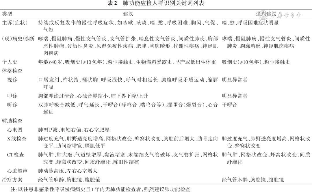
问题1:肺功能检查应检人群如何识别?
【推荐意见1】通过明确肺功能检查应检人群的关键词(表2),帮助临床医师加强对肺功能异常高风险人群的识别能力,从而提高肺功能检查率[1,6,16,18,44-48]。肺功能应检人群可通过电子信息系统自动识别门诊或住院病历关键词,弹窗提示。(强推荐)
· 390 ·
中华结核和呼吸杂志2026年4月第49卷第4期 Chin J Tuberc Respir Dis, April 2026, Vol. 49, No. 4
【推荐意见7】
(1) 不同品牌的肺功能仪检查报告格式应当能够与医院信息系统(HIS)和影像信息系统(PACS)、实验室信息系统(LIS)等实现无缝对接、数据转录。(强推荐)
(2)信息系统接入标准及转换要求:建议肺功能报告电子化遵循统一且规范的格式纳入HIS[37],包含受检者的基本资料、检查的指标、数据、图形以及检查操作者或审核医师的电子签名[56]。如暂未能实现数字化的对接,可采用检查报告的图片或PDF格式与医院PACS系统对接录入。(强推荐)
问题8:如何建立医疗机构内标准肺功能数据平台?
【推荐意见8】
(1)院内整合各科室的肺功能检查数据,确保检查数据能够实时、准确地上传至HIS系统,记录肺功能检查方法类型、次数、结果等,建立统一标准的肺功能数据库,实现数据的集中管理和分析。(强推荐)
(2)平台应具备基本的数据存储、查询、统计分析、质量控制等功能,建议在电子化报告中嵌入统一的质量评估分级系统,有助于临床医师快速了解检查质量,判断结果的可靠性。(强推荐)
(3)应将确诊的呼吸慢病患者纳入平台管理,或在患者管理平台加入肺功能信息,为临床研究和医院管理提供支持,实现患者肺功能数据的可视化管理[38,57-58]。(强推荐)
问题9:如何基于云计算搭建数据共享平台?
【推荐意见9】应制定肺功能数据上传区域云端的标准和规范,确保数据安全和患者隐私信息不被泄露;通过区域云端存储,实现区域内医疗机构间的肺功能数据共享,为分级诊疗和远程医疗提供数据支撑;应对受试者信息进行脱敏处理后再上传至区域分中心;区域分中心承担报告审核、修订报告结论以及质控评级的工作;云数据总中心则汇总各分中心的质控数据,定期对分中心的质控报告
进行评价和抽检$^{[58]}$。(强推荐)
问题10:如何提高肺功能检查的质量控制和结果判读?
【推荐意见10】
(1)肺功能检查的质量控制和结果判读应当由受过肺功能检查专业培训并取得相关资质的医师或技师承担。此外,可借助AI实现肺功能检查结果质控和解读,促进医疗资源的优化配置和提高效率。(强推荐)
(2)AI辅助肺功能检查的实施方式:由肺功能检查设备厂家将AI肺功能系统置入仪器或云平台;通过AI深度学习等模型自动识别曲线异常,智能判读肺功能检查结果的可接受性、可重复性和质量评级,更准确区分肺通气功能障碍类型,提高诊断的准确性和效率,实现异常人群的高效自动识别[59]。(强推荐)
解释说明:肺功能检查的质量控制和结果判读当前主要依赖人工评估,效率低且易受主观因素影响,需由经培训有资质的人员实施。引入AI技术可实现对肺功能检查数据的自动质控和辅助判读,提升肺功能检查效率和质量[66]。
基于大量标准肺功能数据的AI深度学习模型可对肺功能检查过程中的数据采集、曲线质量进行实时评估,对不符合质量标准的检查结果及时预警并要求重检[61]。对可接受的肺功能检查结果进一步判断是否存在异常,并评估肺功能损伤的类型和程度,智能生成实时警告、患者指导和辅助判读等,提高肺功能检查结果的质控能力和判读效率。
问题11:肺功能室设备及人员如何配置?
【推荐意见11】
(1)场地配置:医院需设立肺功能检查室,确保空间布局合理,并具备通风条件和温湿度调控装置,达到院感防控要求并为患者提供舒适的检查环境。(强推荐)
(2)硬件配置:按照所在医疗机构需求配备肺 功能检查设备;设备符合行业技术规范,并在使用 中进行定期校准和维护,建立完善的设备管理制度 档案[38]。(强推荐)
(3)人员配置:根据医疗机构设备和工作量合理配备专职技术人员。人员应经过相关培训并通过考核[42]。(强推荐)
问题12:如何改善就医体验,提升肺功能检查效率?
中华结核和呼吸杂志2026年4月第49卷第4期 Chin J Tuberc Respir Dis, April 2026, Vol. 49, No. 4
· 393 ·
【推荐意见12】
(1)可通过设置肺功能检查自助服务终端或使用手机APP等方式,帮助快速完成挂号、缴费、预约、报到、查看报告等流程,改善肺功能检查就医体验、减少排队等待时间、提升效率。(强推荐)
(2)播放肺功能检查科普视频,可以直观地让肺功能检查者学习肺功能检查的意义、了解检查过程及结果,配合检查方法及关注相应的注意事项。(强推荐)
(3)采用增强现实技术(如AR眼镜)等措施有助于提升快速学习和检查效率[62]。为患者提供代入式的互动体验,增强患者的参与感和理解度,有助于提高肺功能检查的开展率和效率。(强推荐)
问题13:呼吸相关学科开展肺功能检查时,如何针对不同科室设定目标检查开展率?
[推荐意见13]建议除呼吸和胸外科以外,其他相关临床科室开具的肺功能检查数量总和应达到全院肺功能检查总数的一定占比,如30%以上。(强推荐)
问题14:如何设定肺功能检查考核机制?
[推荐意见14]需建立肺功能检查医师和技师综合考核标准,考核维度需包括检查开展及完成的数量和质量。对不达标的情况进行分析和修正,如人员再培训和再考核、补充配备人员和仪器设备,优化绩效评价等。(强推荐)
三、总结与展望
本共识针对当前肺功能临床应用过程中面临的实际问题,围绕优化肺功能检查路径与管理目标,从设备、流程、管理、协作四个维度提出了系统性解决方案及可行性建议,并基于上述推荐意见规定了相应的分级医院建设标准。
我国肺功能检查的标准化管理路径和协作机制尚处于初步探索阶段,目前仍缺乏多中心大样本的临床研究证据,当前共识主要基于专家临床经验形成推荐意见。未来,在肺功能规范化路径推行过程中,需根据国家政策与技术进步动态更新评价标准,开展更多高质量临床研究,为推荐意见提供更充分的循证医学证据;同时需不断完善信息化质控与管理体系,发展多学科协作,基于临床反馈,进一步明确肺功能应检人群特征及各科室标准化实施路径;并通过健全组织保障与激励机制,全面提升肺功能检查管理水平。通过结合不断积累的循证证据和实践反馈,持续修订肺功能检查路径与管理标准,从而为提升我国呼吸系统疾病的整体防治水
平提供坚实支撑。
专家组名单
撰写组(以姓氏拼音排序):陈芳(浙江省中医院);高怡(广州医科大学附属第一医院);郭健(上海市肺科医院);韩伟(康复大学青岛医院);梁斌苗(四川大学华西医院);刘先胜(山西白求恩医院);刘晓菊(兰州大学第一医院);逯勇(首都医科大学附属北京朝阳医院);沈宁(北京大学第三医院);宋元林(复旦大学附属中山医院);孙培莉(南京医科大学第一附属医院);张旭华(宁夏医科大学总医院);张云辉(云南省第一人民医院);郑劲平(广州医科大学附属第一医院);周明娟(广东省中医院)
循证证据组(以姓氏拼音排序): 戴然(上海交通大学医学院附属瑞金医院); 郭健(上海市肺科医院); 逯勇(首都医科大学附属北京朝阳医院); 马千里(重庆松山医院); 孙培莉(南京医科大学第一附属医院)
审阅组(以姓氏拼音排序):包海荣(兰州大学第一医院);薄建萍(山西医科大学第二医院);蔡珊(中南大学湘雅二医院);操敏(首都医科大学附属北京胸科医院);曹璐(解放军总医院第一医学中心);陈芳(浙江省中医院);陈海娟(西安交通大学第二附属医院);陈淑云(南昌大学第一附属医院);陈思敏(广西医科大学第一附属医院);陈亚红(北京大学第三医院);程克斌(上海市肺科医院);戴然(上海交通大学医学院附属瑞金医院);迪丽努尔(厦门市海沧医院);高怡(广州医科大学附属第一医院);龚颖(复旦大学附属中山医院);巩晓红(河北医科大学第四医院);郭健(上海市肺科医院);韩文(山西白求恩医院);黄敏(山西医科大学第二医院);贲慧英(新疆维吾尔自治区人民医院);孔玉红(内蒙古包钢医院);李飞(苏州大学附属第一医院);李丽(复旦大学附属中山医院);梁斌苗(四川大学华西医院);刘银芳(白银市第一人民医院);逯勇(首都医科大学附属北京朝阳医院);马千里(重庆松山医院);苗丽君(郑州大学第一附属医院);倪望(华中科技大学同济医学院附属同济医院);彭毅(华中科技大学同济医学院附属协和医院);秦岭(中南大学湘雅医院);阚呈立(北京大学第一医院);沈北兰(广州医科大学附属第一医院);沈瑶(上海市浦东医院);孙培莉(南京医科大学第一附属医院);所鸿(内蒙古医科大学附属医院);田新瑞(山西医科大学第二医院);王得翔(山东大学齐鲁医院);王惠妩(新疆医科大学第一附属医院);王静(山东省公立医院);谢燕清(广州医科大学附属第一医院);熊维宁(上海交通大学医学院附属第九人民医院);杨卫江(新疆医科大学第四附属医院);杨翼萌(北京医院);姚一旆(浙江大学医学院附属第一医院);云春梅(内蒙古自治区人民医院);张晓雷(中日友好医院);张旭华(宁夏医科大学总医院);赵桂华(河南省人民医院);赵海金(南方医科大学南方医院);赵伟(新疆医科大学第一附属医院);赵伟业(青岛市市立医院);周明娟(广东省中医院);周宁(天津医科大学总医院);邹义敏(浙江大学医学院附属第二医院)
利益冲突所有作者声明无利益冲突
参考文献
[1]中华医学会呼吸病学分会慢性阻塞性肺疾病学组,中国医师协会呼吸医师分会慢性阻塞性肺疾病工作委员会.慢性
· 394 ·
中华结核和呼吸杂志2026年4月第49卷第4期 Chin J Tuberc Respir Dis, April 2026, Vol. 49, No. 4
阻塞性肺疾病诊治指南(2021年修订版)[J].中华结核和呼吸杂志,2021,44(3):170-205.DOI:10.3760/cma.j.cn112147-20210109-00031.
[2] Global Initiative for Chronic Obstructive Lung Disease. Global strategy for prevention, diagnosis and management of COPD: 2025 Report[EB/OL]. [2025-05-23]. https://goldcopd.org/2025-gold-report/.
[3] Global Initiative for Asthma. The global strategy for asthma management and prevention [EB/OL]. [2025-05-06]. https://ginasthma.org/reports/.
[4]中华医学会呼吸病学分会哮喘学组.支气管哮喘防治指南(2020年版)[J].中华结核和呼吸杂志,2020,43(12):1023-1048.DOI:10.3760/cma.j.cn112147-20200618-00721.
[5]中华医学会呼吸病学分会,中国医师协会呼吸医师分会.间质性肺疾病多学科讨论规范中国专家共识[J].中华结核和呼吸杂志,2023,46(12):1176-1188.DOI:10.3760/cma.j.cn112147-20230726-00030.
[6]中华医学会,中华医学会杂志社,中华医学会全科医学分会,等.中国咳嗽基层诊疗与管理指南(2024年)[J].中华全科医师杂志,2024,23(8):793-812.DOI:10.3760/cma.j.cn114798-20240326-00177.
[7] Simons SO, Elliott A, Sastry M, et al. Chronic obstructive pulmonary disease and atrial fibrillation: an interdisciplinary perspective[J]. Eur Heart J, 2021, 42(5): 532-540. DOI: 10.1093/eurheartj/ehaa822.
[8] Buch P, Friberg J, Scharling H, et al. Reduced lung function and risk of atrial fibrillation in the Copenhagen City Heart Study[J]. Eur Respir J, 2003, 21(6): 1012-1016. DOI: 10.1183/09031936.03.00051502.
[9] Brunelli A, Refai MA, Salati M, et al. Carbon monoxide lung diffusion capacity improves risk stratification in patients without airflow limitation: evidence for systematic measurement before lung resection[]]. Eur J Cardiothorac Surg, 2006, 29(4):567-570. DOI: 10.1016/j. ejcts.2006.01.014.
[10] Win T, Laroche CM, Groves AM, et al. Use of quantitative lung scintigraphy to predict postoperative pulmonary function in lung cancer patients undergoing lobectomy[J]. Ann Thorac Surg, 2004, 78(4):1215-1218. DOI: 10.1016/j. athoracsur.2004.04.010.
[11] Larsen KR, Svendsen UG, Milman N, et al. Cardiopulmonary function at rest and during exercise after resection for bronchial carcinoma[]]. Ann Thorac Surg, 1997, 64(4): 960-964. DOI: 10.1016/s0003-4975(97)00635-8.
[12] Powers BK, Ponder HL, Findley R, et al. Enhanced recovery after surgery (ERAS(®))] society abdominal and thoracic surgery recommendations: a systematic review and comparison of guidelines for perioperative and pharmacotherapy core items[J]. World J Surg, 2024, 48(3): 509-523. DOI: 10.1002/wjs.12101.
[13]支修益,刘伦旭.中国胸外科围手术期气道管理指南(2020版)[J].中国胸心血管外科临床杂志,2021,28(3):251-262.DOI:10.7507/1007-4848.202012090.
[14] Liang H, Che G, Cui F, et al. Multidisciplinary international expert consensus on perioperative airway management [J]. Transl Lung Cancer Res, 2025, 14(4):1042-1060. DOI: 10.21037/tlcr-2025-273.
[15] Dankert A, Dohrmann T, Löser B, et al. Pulmonary function tests for the prediction of postoperative pulmonary complications[J]. Dtsch Arztebl Int, 2022, 119(7):99-106. DOI: 10.3238/arztebl.m2022.0074.
[16] Ntima NO, Lumb AB. Pulmonary function tests in anaesthetic practice[]]. BJA Educ, 2019, 19(7): 206-211. DOI: 10.1016/j.bjae.2019.02.001.
[17] Chandler D, Mosieri C, Kallurkar A, et al. Perioperative strategies for the reduction of postoperative pulmonary complications[J]. Best Pract Res Clin Anaesthesiol, 2020, 34(2):153-166. DOI: 10.1016/j.bpa.2020.04.011.
[18] Wang C, Xu J, Yang L, et al. Prevalence and risk factors of chronic obstructive pulmonary disease in China (the China Pulmonary Health [CPH] study): a national cross-sectional study[J]. Lancet, 2018, 391(10131): 1706-1717. DOI: 10.1016/S0140-6736(18)30841-9.
[19]中国老年学和老年医学学会.老年慢性阻塞性肺疾病管理指南[J].中西医结合研究,2023,15(3):154-164.DOI:10.3969/j.issn.1674-4616.2023.03.003.
[20] Chang AB, Grimwood K, Boyd J, et al. Management of children and adolescents with bronchiectasis: summary of the ERS clinical practice guideline[J]. Breathe (Sheff), 2021, 17(3): 210105. DOI: 10.1183/20734735.0105-2021.
[21]周梦雪.肺功能检查在小儿外科术前评估中的应用研究进展[].临床小儿外科杂志,2021,20(4):382-387.DOI:10.12260/lcxewkzz.2021.04.015.
[22]王雪瑶,潘伟华,王伟鹏,等.先天性膈疝患儿术后肺功能评估及影响因素分析[1].上海交通大学学报(医学版),2021,(9):1169-1174.DOI:10.3969/j.issn.1674-8115.2021.09.006.
[23] Yazdanpanah L, Shidfar F, Moosavi AJ, et al. Energy and protein intake and its relationship with pulmonary function in chronic obstructive pulmonary disease (COPD) patients[]. Acta Med Iran, 2010, 48(6):374-379.
[24]中国医师协会呼吸医师分会,中华医学会呼吸病学分会,中国康复医学会呼吸康复专业委员会,等.中国慢性呼吸道疾病呼吸康复管理指南(2021年)[J].中华健康管理学杂志,2021,15(6):521-538.DOI:10.3760/cma.j.cn115624-20211011-00595.
[25]中国疾病预防控制中心慢性非传染性疾病预防控制中心.中国居民慢性阻塞性肺疾病监测报告(2019-2020)[M].北京:人民卫生出版社,2025.
[26] 孙永昌,张静,陈亚红,等.呼吸专科医师慢性阻塞性肺疾病诊断和评估工具应用状况调查[].中华结核和呼吸杂志,2022, 45(4): 362-367. DOI: 10.3760/cma. j.cn112147-20211119-00818.
[27]文富强,申永春,陈磊.肺功能检查在中国慢性阻塞性肺疾病患者中的实践:挑战与对策[J].中华结核和呼吸杂志,2017,40(12):891-893.DOI:10.3760/cma.j.issn.1001-0939.2017.12.004.
[28]中华医学会呼吸病学分会肺功能专业组.肺功能检查指南(第一部分)——概述及一般要求[J].中华结核和呼吸杂志,2014, 37(6): 402-405. DOI: 10.3760/cma.j.issn.1001-0939.2014.06.002.
[29]中华医学会呼吸病学分会肺功能专业组.肺功能检查指南(第二部分)——肺量计检查[J].中华结核和呼吸杂志,2014,37(7):481-486.DOI:10.3760/cma.j.issn.1001-0939.2014.07.001.
[30] 中华医学会呼吸病学分会肺功能专业组.肺功能检查指南(第三部分)--组织胺和乙酰甲胆碱支气管激发试验[J].中华结核和呼吸杂志,2014,37(8):566-571.DOI:10.3760/cma.j.issn.1001-0939.2014.08.003.
[31]中华医学会呼吸病学分会肺功能专业组.肺功能检查指南(第四部分)——支气管舒张试验[J].中华结核和呼吸杂志,2014, 37(9): 655-658. DOI: 10.3760/cma.j.issn.1001-0939.2014.09.007.
[32]中华医学会呼吸病学分会肺功能专业组.肺功能检查指南--
中华结核和呼吸杂志2026年4月第49卷第4期 Chin J Tuberc Respir Dis, April 2026, Vol. 49, No. 4
· 395 ·
肺容量检查[J].中华结核和呼吸杂志,2015,38(4):255-260.DOI:10.3760/cma.j.issn.1001-0939.2015.04.005.
[33]中华医学会呼吸病学分会肺功能专业组.肺功能检查指南-肺弥散功能检查[].中华结核和呼吸杂志,2015,38(3):164-169.DOI:10.3760/cma.j.issn.1001-0939.2015.03.003.
[34] 中华医学会呼吸病学分会肺功能专业组.肺功能检查指南——体积描记法肺容量和气道阻力检查[J].中华结核和呼吸杂志,2015,38(5):342-347.DOI:10.3760/cma.j.issn.1001-0939.2015.05.006.
[35]中华医学会呼吸病学分会肺功能专业组.肺功能检查指南-呼气峰值流量及其变异率检查[J].中华结核和呼吸杂志,2017,40(6):426-430.DOI:10.3760/cma.j.issn.1001-0939.2017.06.006.
[36]中华医学会,中华医学会杂志社,中华医学会全科医学分会,等.中国常规肺功能检查基层指南(2024年)[J].中华全科医师杂志,2025,24(2):121-137.DOI:10.3760/cma.j.cn114798-20240618-00555.
[37] 中国呼吸医师协会肺功能与临床呼吸生理工作委员会,中华医学会呼吸病学分会呼吸治疗学组.肺功能检查报告规范——肺量计检查、支气管舒张试验、支气管激发试验.].中华医学杂志,2019,99(22):1681-1691.DOI:10.3760/cma.j.issn.0376-2491.2019.22.001.
[38]国家呼吸系统疾病临床医学研究中心,呼吸疾病国家重点实验室,国家呼吸医学中心.肺功能检查管理规范[M].北京:人民卫生出版社,2020.
[39]中国医师协会呼吸医师分会肺功能与临床呼吸生理工作委员会,中华医学会呼吸病学分会肺功能学组,中国老年医学会呼吸分会肺功能学组.肺功能检查技术规范——脉冲振荡技术检查[J].中华结核和呼吸杂志,2022,[10]:960-969.DOI:10.3760/cma.j.cn112147-20220127-00080.
[40] 中国医师协会呼吸医师分会肺功能与临床呼吸生理工作委员会,中华医学会呼吸病学分会,中国老年医学会呼吸分会肺功能学组.乙酰甲胆碱(氯醋甲胆碱)支气管激发试验技术规范(2023年版)[J].中华结核和呼吸杂志,2024,47(2):101-119.DOI:10.3760/cma.j.cn112147-20231019-00247.
[41]中华医学会呼吸病学分会肺功能学组.便携式肺功能仪原理、质控及临床应用的中国专家共识[J].中华结核和呼吸杂志,2022,[10]:970-979.DOI:10.3760/cma.j.cn112147-20220302-00167.
[42] 安嘉颖,虞欣欣,沈北兰,等.中国肺功能检查临床应用与质量控制规范化培训考核认证方案概述[J].中国实用内科杂志,2021,(9):821-824.DOI:10.19538/j.nk2021090115.
[43] 高怡,郑劲平.开展肺功能规范化培训,助力慢性呼吸系统疾病综合防控[J].中国实用内科杂志,2019,39(5):481-484.DOI:10.19538/j.nk2019050101.
[44] Evans SE, Scanlon PD. Current practice in pulmonary function testing[J]. Mayo Clin Proc, 2003, 78(6):758-763; quiz 763. DOI: 10.4065/78.6.758.
[45] Ranu H, Wilde M, Madden B. Pulmonary function tests[J]. Ulster Med J, 2011, 80(2):84-90.
[46] Graham BL, Steenbruggen I, Miller MR, et al. Standardization of spirometry 2019 update. An official American thoracic society and European respiratory society technical statement [J]. Am J Respir Crit Care Med, 2019, 200(8):e70-e88. DOI: 10.1164/rccm.201908-1590ST.
[47] 中华医学会呼吸病学分会肺功能学组.成人肺功能检查技术进展及临床应用推荐指南(2025版)[J].中华肺部疾病杂志(电子版),2025,18(2):197-212.DOI:10.3877/cma.j.issn.1674-6902.2025.02.001.
[48] Brunelli A, Kim AW, Berger KI, et al. Physiologic evaluation of the patient with lung cancer being considered for resectional surgery: Diagnosis and management of lung cancer, 3rd ed: American College of Chest Physicians evidence-based clinical practice guidelines[J]. Chest, 2013, 143(5 Suppl): e166S-e190S. DOI: 10.1378/chest.12-2395.
[49] Zhou YM, Chen SY, Tian J, et al. Development and validation of a chronic obstructive pulmonary disease screening questionnaire in China[J]. Int J Tuberc Lung Dis, 2013, 17(12):1645-1651. DOI: 10.5588/ijtld.12.0995.
[50] Yawn BP, Mapel DW, Mannino DM, et al. Development of the Lung Function Questionnaire (LFQ) to identify airflow obstruction[J]. Int J Chron Obstruct Pulmon Dis, 2010, 5: 1-10.
[51]郑劲平.肺功能检查临床意义和诊断思路[J].中国实用内科杂志,2012,32(8):569-574.
[52]宋元林,盖晓燕.像量血压一样测肺功能,如何评价和改善肺功能质控?[J].中国全科医学,2023,26(5):539.
[53]陆海英、袁泉、徐隽、等.肺功能监测和CT肺功能成像检查结果的相关性分析[J].现代生物医学进展,2016,16(34):6750-6753.DOI:10.13241/j.cnki.pmb.2016.34.039.
[54] Stolz D, Mkorombindo T, Schumann DM, et al. Towards the elimination of chronic obstructive pulmonary disease: a Lancet Commission[]]. Lancet, 2022, 400(10356): 921-972. DOI: 10.1016/S0140-6736(22)01273-9.
[55] Sharkey MJ, Checkley EW, Swift A J. Applications of artificial intelligence in computed tomography imaging for phenotyping pulmonary hypertension[J]. Current Opinion in Pulmonary Medicine, 2024, 30(5): 464-472. DOI: 10.1097/MCP.0000000000001103.
[56]顾明豪,郑蕴欣,王愿颖.基于临床需求的肺功能信息化系统构建与应用评估[J].中国医学装备,2023,20(8):106-110.DOI:10.3969/J.ISSN.1672-8270.2023.08.023.
[57]赖莉芬,常晓红,韩志海,等.肺功能检查室数字化管理系统设计与实现[J].医疗卫生装备,2017,38(9):51-53,57.DOI:10.7687/j.issn1003-8868.2017.09.051.
[58] 司超增,胡宇,夏沁芳,等.肺功能检查全流程数智化管理系统的构建及临床应用[J].中国医疗设备,2025,40(6):69-74,81.DOI:10.3969/j.issn.1674-1633.20241559.
[59] Wang Y, Li Y, Chen W, et al. Deep learning for spirometry quality assurance with spirometric indices and curves[J]. Respir Res, 2022, 23(1): 98. DOI: 10.1186/s12931-022-02014-9.
[60] Al-Anazi S, Al-Omari A, Alanazi S, et al. Artificial intelligence in respiratory care: Current scenario and future perspective[J]. Ann Thorac Med, 2024, 19(2): 117-130. DOI: 10.4103/atm.atm_192_23.
[61] Gompelmann D, Gysan MR, Desbordes P, et al. Al-powered evaluation of lung function for diagnosis of interstitial lung disease[J]. Thorax, 2025, 80(7): 445-450. DOI: 10.1136/thorax-2024-221537.
[62] Chen Y, Zhang Y, Long X, et al. Effectiveness of virtual reality-complemented pulmonary rehabilitation on lung function, exercise capacity, dyspnea, and health status in chronic obstructive pulmonary disease: systematic review and meta-analysis[J]. J Med Internet Res, 2025, 27: e64742. DOI: 10.2196/64742.
| 科室 | 肺功能检查的应用价值 |
|---|---|
| 呼吸科[1-6] | 诊断慢阻肺病,诊断哮喘,慢性咳嗽病因诊断,评估间质性肺疾病进展,评估多种呼吸疾病严重程度,慢病监测及管理,新药开发及上市评价 |
| 心内科[7-8] | 与呼吸系统疾病进行鉴别诊断,识别是否合并慢阻肺病、哮喘等呼吸系统疾病,临床预后评估,指导心肺康复 |
| 胸外科/腹部外科[9-16] | 术前预测围手术期肺部并发症风险,术前指导制定手术方案,术后肺功能监测,术后指导呼吸康复 |
| 麻醉科[16-17] | 术前评估、术中管理和术后监测 |
| 老年病科[18-19] | 筛查慢阻肺病患者,识别慢阻肺病高危人群,早期干预 |
| 儿科[3,20-22] | 儿科呼吸疾病的诊断和评估,先天肺发育异常儿童的评估和随访 |
| 健康管理中心/体检中心 | 早期识别呼吸生理异常,筛查慢阻肺病等呼吸系统疾病患者 |
| 其他学科[4-23-24] | 评估呼吸系统合并症,辅助进行整体健康评估,指导呼吸康复评定,监测治疗疗效和安全性,评估疾病进展和预后 |
| 类型 | 建议 | 强烈建议 |
|---|---|---|
| 主诉(症状) | 持续或反复发作的慢性呼吸症状,如咳嗽、咳痰、喘、憋、呼吸困难、胸闷、气促、气短 | 喘、憋、呼吸困难症状明显 |
| (现)病史/诊断 | 哮喘、慢阻肺病、慢性支气管炎、支气管扩张、喘息性支气管炎、间质性肺炎、胸部恶性肿瘤、过敏性鼻炎、风湿免疫性疾病、肥胖、胸廓畸形、代谢性疾病、神经肌肉疾病 | 哮喘、慢阻肺病、慢性支气管炎、间质性肺炎、胸廓畸形、神经肌肉疾病 |
| 个人史 | 年龄≥40岁,吸烟史(≥10包年)、粉尘接触史、生物燃料暴露史、早产或低出生体重 | 吸烟史(≥10包年)、粉尘接触史 |
| 体格检查 | ||
| 视诊 | 口唇发绀、杆状指、桶状胸、呼吸浅快、呼气时相延长、胸腹呼吸矛盾运动、缩唇呼吸 | 明显异常者 |
| 叩诊 | 胸部叩诊过清音、心浊音界缩小、肺下界下降/上升 | 明显异常者 |
| 听诊 | 双肺呼吸音减低、呼气延长、干啰音(哮鸣音、喘鸣音等)、湿啰音(爆裂音)、心音遥远 | 干啰音 |
| 辅助检查 | ||
| 心电图 | 肺型P波、电轴右偏、右心室肥厚 | |
| X线检查 | 肺过度充气,肺野透亮度增高,网格状改变、蜂窝状改变、胸腔前后增大,肋骨走向变平,肋间隙增宽、膈肌低平 | 肺过度充气、肺野透亮度增高、网格状改变、蜂窝状改变 |
| CT检查 | 肺气肿、肺大疱、气道壁增厚、黏液堵塞、末端细支气管破坏、支气管扩张、网格状改变、蜂窝状改变、间质纤维化、陈旧性结核 | 肺气肿、网格状改变、蜂窝状改变、间质纤维化 |
| 心脏超声 | 肺动脉高压,左右心室增大 | |
| 治疗方案 | 经气管麻醉、胸腔镜、腹腔镜 | 经气管麻醉、胸腔镜、腹腔镜 |
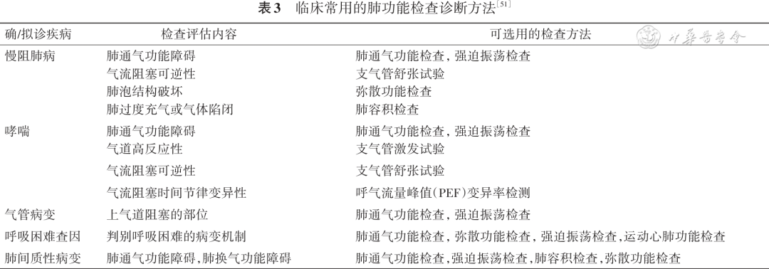
| 确/拟诊疾病 | 检查评估内容 | 可选用的检查方法 |
|---|---|---|
| 慢阻肺病 | 肺通气功能障碍 | 肺通气功能检查,强迫振荡检查 |
| 慢阻肺病 | 气流阻塞可逆性 | 支气管舒张试验 |
| 慢阻肺病 | 肺泡结构破坏 | 弥散功能检查 |
| 慢阻肺病 | 肺过度充气或气体陷闭 | 肺容积检查 |
| 哮喘 | 肺通气功能障碍 | 肺通气功能检查,强迫振荡检查 |
| 哮喘 | 气道高反应性 | 支气管激发试验 |
| 哮喘 | 气流阻塞可逆性 | 支气管舒张试验 |
| 哮喘 | 气流阻塞时间节律变异性 | 呼气流量峰值(PEF)变异率检测 |
| 气管病变 | 上气道阻塞的部位 | 肺通气功能检查,强迫振荡检查 |
| 呼吸困难查因 | 判别呼吸困难的病变机制 | 肺通气功能检查,弥散功能检查,强迫振荡检查,运动心肺功能检查 |
| 肺间质性病变 | 肺通气功能障碍,肺换气功能障碍 | 肺通气功能检查,强迫振荡检查,肺容积检查,弥散功能检查 |


相关指南
引用 / 出处
中国医师协会呼吸医师分会肺功能与临床呼吸生理工作组. 优化肺功能检查多学科临床路径与管理模式专家共识[J]. 中华结核和呼吸杂志, 2026.