儿童淋巴结肿大临床诊治专家共识(2026)
参考文献
[1] Yenilmez E, Verdi Y, Ilbak A, et al. Demographic, clinical and laboratory characteristics for differential diagnosis of peripheral lymphadenopathy (LAP) and the etiologic distribution of LAP in adults; a multicenter; nested case-control study including 1401 patients from Turkey [J]. Intern Emerg Med, 2021, 16(8): 2139-2153. DOI: 10.1007/s11739-021-02683-2.
[2] Schwab TC, Perrig L, Göller PC, et al. Targeted next-generation sequencing to diagnose drug-resistant tuberculosis: a systematic review and meta-analysis[J]. Lancet Infect Dis, 2024, 24(10):1162-1176. DOI: 10.1016/S1473-3099(24)00263-9.
[3] Deosthali A, Donches K, DelVecchio M, et al. Etiologies of pediatric cervical lymphadenopathy: a systematic review of 2687 subjects[J]. Glob Pediatr Health, 2019, 6: 2333794X19865440. DOI: 10.1177/2333794X19865440.
[4] 陈耀龙,杨克虎,王小钦等.中国制订/修订临床诊疗指南的指导原则(2022版)[J].中华医学杂志,2022,102(10):697-703. DOI:10.3760/cma.j.cn112137-20211228-02911.
[5] Long SS, Prober CG, Fischer M, et al. Principles and practice of pediatric infectious diseases[M]. 6th ed. Amsterdam: Elsevier, 2022. DOI: 10.1016/C2019-0-00075-1
[6] National Collaborating Centre For Cancer (UK). Suspected cancer: recognition and referral[M]. London: National Institute for Health and Care Excellence (NICE), 2015.
[7] Oxford Centre for Evidence-Based Medicine. The oxford levels of evidence 2 [EB/OL]. 2009[2025-06-01]. https://www. cebm. ox. ac. uk/resources/levels-of-evidence/ocebm-levels-of-evidence.
[8] Alves Rosa J, Calle-Toro JS, Kidd M, et al. Normal head and neck lymph nodes in the paediatric population[J]. Clin Radiol, 2021, 76(4): 315. e-315. e7. DOI: 10.1016/j.crad.2020.12.020.
[9] Dogan MS, Koc G, Doganay S, et al. Computed tomography depiction of normal inguinal lymph nodes in children[J]. Folia Morphol (Warsz), 2020, 79(4): 799-804. DOI: 10.5603/FM/a2020.0005.
[10] de Jong PA, Nievelstein RJ. Normal mediastinal and hilar lymph nodes in children on multi-detector row chest computed tomography[J]. Eur Radiol, 2012, 22(2): 318-321. DOI: 10.1007/s00330-011-2253-9.
[11] Karmazyn B, Werner EA, Rejaie B, et al. Mesenteric lymph nodes in children: what is normal? [J]. Pediatr Radiol, 2005, 35(8):774-777. DOI: 10.1007/s00247-005-1462-2.
[12] Karmazyn B, Supakul N, Lehnert SJ, et al. Retroperitoneal and pelvic lymph nodes in children: what is normal? [J]. AJR Am J Roentgenol, 2020, 214(6): 1384-1388. DOI: 10.2214/AJR.19.22316.
[13] Ye F, Ling W, Wu Q, et al. Porta hepatis lymph nodes on US: not only identify biliary atresia but also predict outcomes after Kasai portoenterostomy surgery[J]. Insights Imaging, 2024, 15(1):154. DOI: 10.1186/s13244-024-01735-3.
[14] Wang J, Pei G, Yan J, et al. Unexplained cervical
中华儿科杂志 2026年1月第64卷第1期 Chin J Pediatr, January 2026, Vol. 64, No. 1
· 41 ·
lymphadenopathy in children: predictive factors for malignancy[J]. J Pediatr Surg, 2010, 45(4):784-788. DOI: 10.1016/j.jpedsurg.2009.08.018. [15] Yaris N, Cakir M, Sözen E, et al. Analysis of children with peripheral lymphadenopathy[J]. Clin Pediatr (Phila), 2006, 45(6):544-549. DOI: 10.1177/0009922806290609. [16] Haimi-Cohen Y, Elron E, Oz-Alcalay L, et al. Risk factors for malignancy in pediatric subacute/chronic focal craniocervical lymphadenopathy[J]. Front Pediatr, 2024, 12:1466116. DOI: 10.3389/fped.2024.1466116. [17] Zhu J, Ma J, Union for China Lymphoma Investigators of Chinese Society of Clinical Oncology, et al. Chinese Society of Clinical Oncology (CSCO) diagnosis and treatment guidelines for malignant lymphoma 2021 (English version)[J]. Chin J Cancer Res, 2021, 33(3):289-301. DOI: 10.21147/j.issn.1000-9604.2021.03.01. [18] Linabery AM, Erhardt EB, Richardson MR, et al. Family history of cancer and risk of pediatric and adolescent Hodgkin lymphoma: a Children's Oncology Group study [J]. Int J Cancer, 2015, 137(9):2163-2174. DOI: 10.1002/ Otolaryng, 2021, 2022. J. J. J. J. J. J. J. J. J. J. J. J. J. J. J. J. J. J. J. J. J. J. J. J. J. J. J. J. J. J. J. J. J. J. J. J. J. J. J. J. J. J. J. J. J. J. J. J. J. J. J. J. J. J. J. J. J. J. J. J. J. J. J. J. J. J. J. J. J. J. J. J. J. J. J. J. J. J. J. J. J. J. J. J. J. J. J. J. J. J. J. J. J. J. J. J. J. J. J. J. J. J. J. J.
hypermetabolic mediastinal-hilar lymph nodes determined by PET/CT with EBUS-TBNA and calculation of SUVmax cutoff values in differentiation of malignancy [J]. Medicine (Baltimore), 2023, 102(35): e34928. DOI: 10.1097/MD.00000000000034928. [29] Thompson JA, Bertoni D, Decuzzi J, et al. Ultrasound versus fine needle aspiration for the initial evaluation of pediatric cervical lymphadenopathy-a systematic review [J]. Int J Pediatr Otorhinolaryngol, 2023, 166: 111485. DOI: 10.1016/j.ijporl.2023.111485. [30] Kim JW, Baek JV, Lee JY, et al. Pathologic etiology and predictors of malignancy in children with cervical lymphadenopathy [J]. World J Pediatr, 2023, 19(3): 283-287. DOI: 10.1007/s12519-022-00667-6. [31] Ye X, Tucker C, Gardner C. Assessment of the diagnostic accuracy of core needle biopsies in the diagnosis of lymphoma[J]. Blood, 2020, 136 (Supplement 1): 12-13. DOI:10.1182/blood-2020-143306. [32] Rehell M, Atula T, Tapiovaara LK, et al. Complications in lymph node excision in the head and neck area [J]. Acta Otolaryngol, 2022, 142(9-12): 738-742. DOI: 10.1080/00016489.2022.2115551. [33] Ha H, Lee J, Kim DY, et al. Utility and limitations of fine-needle aspiration cytology in the diagnosis of lymphadenopathy [J]. Diagnostics (Basel), 2023, 13(4): 728. DOI: 10.3390/diagnostics13040728. [34] Kwon Y, Lee MK. Diagnostic performance and safety of ultrasound-guided core needle biopsy for diagnosing lymphoma: a systematic review and meta-analysis [J]. Cancer Med, 2025, 14(1):e70414. DOI: 10.1002/cam4.70414. [35] 中国人民解放军总医院第八医学中心结核病医学部,《中国防痨杂志》编辑委员会,中国医疗保健国际交流促进会结核病防治分会.线表淋巴结结核的诊断与治疗专家共识[J].中国防痨杂志,2023,45(6):531-542. DOI: 10.19982/j.issn.1000-6621.20230120. [36] Wu J, Song W, Yan H, et al. Metagenomic next-generation sequencing in detecting pathogens in pediatric oncology patients with suspected bloodstream infections [J]. Pediatr Res, 2024, 95(3): 843-851. DOI: 10.1038/s41390-023-02776-y. [37] Song P, Tian Y, Chen S, et al. A novel method for simultaneous detection of hematological tumors and infectious pathogens by metagenomic next generation sequencing of plasma [J]. Clin Chim Acta, 2024, 557: 117874. DOI: 10.1016/j.cca.2024.117874. [38] Chiappini E, Camaioni A, Benazzo M, et al. Development of an algorithm for the management of cervical lymphadenopathy in children: consensus of the Italian Society of Preventive and Social Pediatrics, jointly with the Italian Society of Pediatric Infectious Diseases and the Italian Society of Pediatric Otorhinolaryngology [J]. Expert Rev Anti Infect Ther, 2015, 13(12): 1557-1567. DOI: 10.1586/14787210.2015.1096777. [39] Sartelli M, Guirao X, Hardcastle TC, et al. 2018 WSES/SIS-E consensus conference: recommendations for the management of skin and soft-tissue infections [J]. World J Emerg Surg, 2018, 13: 58. DOI: 10.1186/s13017-018-0219-9. [40] Miller LG, Daum RS, Creech CB, et al. Clindamycin versus trimethoprim-sulfamethoxazole for uncomplicated skin infections [J]. N Engl J Med, 2015, 372(12): 1093-1103.
· 42 ·
中华儿科杂志2026年1月第64卷第1期 Chin J Pediatr, January 2026, Vol. 64, No. 1
DOI: 10.1056/NEJMoa1403789. [41] Key S, Lee M, Kwok M, et al. Management of paediatric neck abscess with needle aspiration against surgical drainage: systematic review and meta-analysis[J]. Int J<y_109><x_354><y_142><LAYOUT_TEXT>10.1016/j.ijporl.2025.112318. [42] McMullan BJ, Andresen D, Blyth CC, et al. Antibiotic duration and timing of the switch from intravenous to oral route for bacterial infections in children: systematic review and guidelines[J]. Lancet Infect Dis, 2016, 16(8): e139-152. DOI: 10.1016/S1473-3099(16)30024-X. [43] Zeng Y, Ying W, Wang W, et al. Clinical and genetic characteristics of bcg disease in Chinese children: a retrospective study[J]. J Clin Immunol, 2023, 43(4): 756-768. DOI: 10.1007/s10875-022-01422-2. [44] Rodriguez-Takechi SY, Renjifo ME, Medina FJ. Extrapulmonary tuberculosis: pathophysiology and imaging findings[J]. Radiographics, 2019, 39(7): 2023-2037. DOI: 10.1148/rg.2019190109. [45] Kohli M, Schiller I, Dendukuri N, et al. Xpert® MTB/RIF assay for extrapulmonary tuberculosis and rifampicin resistance[J]. Cochrane Database Syst Rev, 2018, 8(8): CD012768. DOI: 10.1002/14651858.CD012768.pub2. [46] Cuello-Garcfa CA, Pérez-Gaxiola G, Jiménez Gutiérrez C. Treating BCG-induced disease in children[J]. Cochrane Database Syst Rev, 2013, 2013(1): CD008300. DOI: 10.1002/14651858.CD008300.pub2. [47] Liu C, Huang M, Liu F, et al. The role of surgical management of BCG vaccine-induced regional suppurative lymphadenitis in children: a 7 years' experience from one medical center[J]. BMC Infect Dis, 2021, 21(1):801. DOI: 10.1186/s12879-021-06531-8. [48] Pang Y, Tan Y, Chen J, et al. Diversity of nontuberculous mycobacteria in eastern and southern China: a cross-sectional study[J]. Eur Respir J, 2017, 49(3): 1601429. DOI: 10.1183/13993003.01429-2016. [49] Zimmermann P, Tebruegge M, Curtis N, et al. The management of non-tuberculous cervicofacial lymphadenitis in children: a systematic review and meta-analysis[J]. J Infect, 2015, 71(1):9-18. DOI: 10.1016/j.jinf.2015.02.010. [50] Prutsky G, Domecq JP, Mori L, et al. Treatment outcomes of human bartonellosis: a systematic review and meta-analysis[J]. J Indt In Infect Dis, 2013, 17(10):e811-819. DOI: 10.1016/j.ijid.2013.02.016.
[51] Augustya P, Bhandari A, Mukund B. Epstein-Barr virus infection in children presenting with acute cervical lymphadenopathy[J]. J Mar Med Soc, 2023, 25(2): 188-192. DOI:10.4303/jmms.jmms29_23. [52] Leung A, Lam JM, Barankin B. Infectious mononucleosis: an updated review[J]. Curr Pediatr Rev, 2024, 20(3): 305-322. DOI:10.2174/1573396320666230801091558. [53] Montoya JG, Liesenfeld O. Toxoplasmosis[J]. Lancet, 2004, 363(9425): 1965-1976. DOI: 10.1016/S0140-6736(4) 16412-X. [54] Woolard MD, Hodgde LM, Jones HP, et al. The upper and lower respiratory tracts differ in their requirement of IFN-gamma and IL-4 in controlling respiratory mycoplasma infection and disease[J]. J Immunol, 2004, 172(11): 6875-6883. DOI: 10.4049/jimmunol.172.11.6875. [55] Yang H, Sun P, Zhou S, et al. Chlamydia psittaci infection induces IFN-I and IL-1B through the cGAS-STING-IRF3/NLRP3 pathway via mitochondrial oxidative stress in human macrophages[J]. Vet Microbiol, 2024, 299:110292. DOI: 10.1016/j.vetmic.2024.110292. [56] Hickman CJ, Stover CK, Joseph SW, et al. Molecular cloning and sequence analysis of a Rickettsia tsutsugamushi 22 kDa antigen containing B-and T-cell epitopes[J]. Microb Pathog, 1991, 11(1):19-31. DOI: 10.1016/0882-4010(91) 90090-w. [57] Kanegaye JT, Van Cott E, Tremoulet AH, et al. Lymph-node-first presentation of Kawasaki disease compared with bacterial cervical adenitis and typical Kawasaki disease[J]. J Pediatr, 2013, 162(6): 1259-1263, 1263.e1-2.DO: 10.1016/j.jpeds.2012.11.064. [58] Liu C, Lin R, Luo M, et al. Clinical characteristics of cervical lymphadenopathy in Kawasaki disease and the generation of related cervical complications: a retrospective cohort study[J]. Eur J Med Res, 2025, 30(1): 295. DOI: 10.1186/s40001-025-02556-w. [59] Xie YP, Xu YW, Li Y, et al. Recurrence of histiocytic necrotizing lymphadenitis in children: a 10-year multicenter retrospective study[J]. J Inflamm Res, 2025, 18:4307-4318. DOI: 10.2147/JIR.S504413. [60] Choi S, Choi HS, Ryu YJ, et al. Characterization of kikuchi-fujimoto disease in children and risk factors associated with its course[J]. J Pediatr, 2023, 260:113515. DOI: 10.1016/j.jpeds.2023.113515.
·编辑部公告·
谨防非法网站冒用本刊名义进行征稿、收费的声明
近期一些网站冒用中华儿科杂志名义征稿,或通过发送“录用通知”收取作者版面费。已有多位作者上当受骗。为维护广大读者和作者的权益及杂志声誉,本刊特此声明。
本刊不收取审稿费,关于退修、录用、缴费等事宜均请务必通过中华医学会远程稿件管理系统(可通过中华医学
会网站首页 http://www.cma.org.cn或本刊网站:http://www.cmaped.org.cn进入进行查询。
本刊联系电话:010-51322412。
本刊编辑部
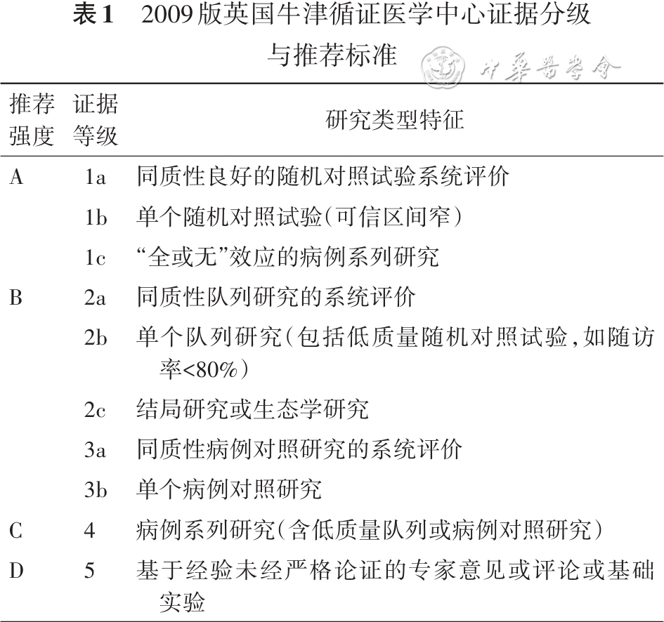
| 推荐 强度 | 证据 等级 | 研究类型特征 |
|---|---|---|
| A | 1a | 同质性良好的随机对照试验系统评价 |
| A | 1b | 单个随机对照试验(可信区间窄) |
| A | 1c | “全或无”效应的病例系列研究 |
| B | 2a | 同质性队列研究的系统评价 |
| B | 2b | 单个队列研究(包括低质量随机对照试验,如随访率<80%) |
| B | 2c | 结局研究或生态学研究 |
| B | 3a | 同质性病例对照研究的系统评价 |
| B | 3b | 单个病例对照研究 |
| C | 4 | 病例系列研究(含低质量队列或病例对照研究) |
| D | 5 | 基于经验未经严格论证的专家意见或评论或基础实验 |
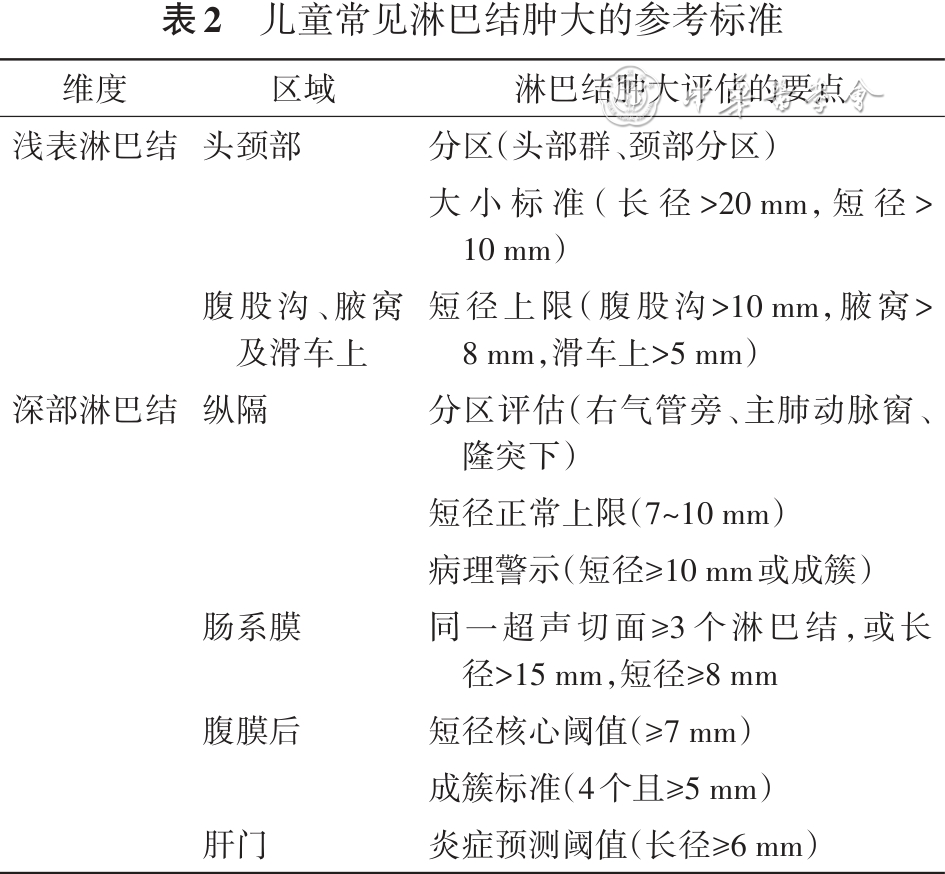
| 维度 | 区域 | 淋巴结肿大评估的要点 |
|---|---|---|
| 浅表淋巴结 | 头颈部 | 分区(头部群、颈部分区) 大小标准(长径>20 mm,短径>10 mm) |
| 短径上限(腹股沟>10 mm,腋窝>8 mm,滑车上>5 mm) | ||
| 深部淋巴结 | 纵隔 | 分区评估(右气管旁、主肺动脉窗、隆突下) 短径正常上限(7~10 mm) 病理警示(短径>10 mm或成簇) 同一超声切面≥3个淋巴结,或长径>15 mm,短径>8 mm 短径核心阈值(≥7 mm) 成簇标准(4个且>5 mm) 炎症预测阈值(长径≥6 mm) |


推荐意见与 GRADE 证据分级
| # | 推荐意见 | 证据等级 | 出处 |
|---|---|---|---|
| 1 | 儿童淋巴结的生理大小因解剖部位而异,个体差异大。当颈部和腹股沟淋巴结长 | — | |
| 2 | 正常淋巴结呈肾形或椭圆形,长径和短径比值(long-axis to short-axis ratio, L/S)通常≥2.0;若比值<2.0,淋巴结趋于圆形,可能存在恶性细胞浸润或其他异常病理状态,需进一步明确病因(证据等级2b,推荐强度B)。 | — | |
| 3 | 儿童淋巴结肿大的评估应基于详尽的病史与全面体格检查,根据淋巴结大小、质地与分布(局部性或全身性)、病史特征(急慢性病程、伴随症状和特殊暴露史),识别潜在的诊断线索,行针对性检查;病因未明时慎用糖皮质激素,必要时活检(证据等级2a,推荐强度B)。 | — | |
| 4 | 推荐超声检查作为初始检查评估形态、结构和血流特征。无法明确诊断淋巴结病变性质时,推荐行磁共振成像或弥散加权磁共振成像,其诊断价值在恶性病变评估中与PET-CT相当(证据等级2b,推荐强度B)。 | — | |
| 5 | 不推荐淋巴结活检作为儿童淋巴结肿大常规的诊疗措施,仅在高度怀疑存在恶性疾病、疑似常规抗感染治疗无效的肉芽肿、伴有全身症状且难以明确诊断时考虑应用(证据等级2b,推荐强度B)。 | — | |
| 6 | 活检方式的选择需综合考虑操作风险、患儿耐受性及对病理诊断的需求。良性或深部病变者优先推荐刺活检,怀疑血液系统恶性疾病时推荐切除活检(证据等级2b,推荐强度B)。 | — | |
| 7 | 淋巴结活检除了病理染色,推荐结合病原涂片、培养、免疫组织化学、分子检测等明确病因。淋巴结组织的病原宏基因组测序可提升复杂感染病原检出率,需结合临床解读结果(证据等级2a,推荐强度B)。 | — | |
| 8 | 急性细菌性淋巴结炎需经验性使用覆盖A组链球菌与金黄色葡萄球菌的抗菌药物治疗。病变位于颌下、颌下或伴牙周疾病、鼻窦炎的年长儿,必要时加用覆盖厌氧菌药物的治疗(证据等级4,推荐强度C)。 | — | |
| 9 | 根据可能的病原体、病情严重程度及当地耐甲氧西林金黄色葡萄球菌等耐药菌流行情况选择经验抗菌药物治疗。治疗48~72h评估效果;若无好转,建议采用静脉抗菌药物治疗或扩大抗菌谱范围。病原体明确后,及时调整为窄谱抗菌药物(证据等级1b,推荐强度A)。 | — | |
| 10 | 若细菌性淋巴结炎局部出现波动感时,应根据脓肿大小、深浅及解剖位置,选择超声引导下穿刺或切开等干预手段(证据等级2a,推荐强度B)。 | — | |
| 11 | 慢性无痛性持续性淋巴结肿大除需排除肿瘤外,还需排查胞内菌、真菌、DNA病毒感染,必要时活检明确感染病原。无典型流行病学史者应排除免疫缺陷(证据等级2a,推荐强度B)。 | — | |
| 12 | 病毒与其他感染性病原是儿童淋巴结肿大的主要原因,EB病毒最为常见。外周无法明确病原伴有全身炎症风暴甚至噬血细胞综合征时,建议淋巴结活检找病原,明确病因去除免疫失调的诱发因素(证据等级2a,推荐强度B)。 | — | |
| 13 | 对于经验性抗感染治疗效果不佳的疑似细菌性颈淋巴结炎患儿,建议动态观察其临床变化,借助心脏冠状动脉超声及实验室检查手段,积极与川崎病,尤其是以淋巴结肿大为首发表现的川崎病进行鉴别(证据等级2b,推荐强度B)。 | — | |
| 14 | 对持续性、多部位或伴有特异性全身症状的淋巴结肿大患儿,尤其伴白细胞下降或者肝功能损伤时,建议扩大鉴别诊断范围,系统排查组织细胞坏死性疾病及SLE等疾病(证据等级3b,推荐强度B)。 | — |
相关指南
- 儿童急性呼吸道感染性疾病合理用药指南 · 呼吸内科
- 儿童A族链球菌咽扁桃体炎临床诊疗专家共识(2025) · 儿科 2025
- 慢性阻塞性肺疾病基层合理用药指南 · 呼吸内科 2020
- 儿童连续性肾替代治疗期间抗菌药物治疗方案调整专家共识(2026) · 感染与传染病 2026
- 急诊成人社区获得性肺炎临床实践指南(2024年版) · 呼吸内科 2024
- 实体肿瘤患者伴发肺炎临床诊疗实践中国专家共识(2024版) · 呼吸内科 2024
引用 / 出处
中华医学会儿科学分会感染学组. 儿童淋巴结肿大临床诊治专家共识(2026)[J]. 中华儿科杂志, 2026.